For individuals dealing with eczema, can incorporating acupuncture into a treatment plan help manage and reduce symptoms?
Contents
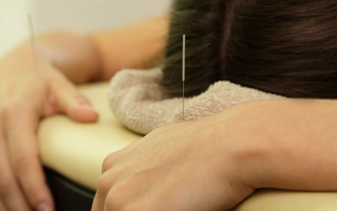
Acupuncture for Eczema
Eczema is a chronic skin disorder that causes intense itching, dry skin, and rashes. Common treatment options for eczema include:
Moisturizers
Topical steroids
Prescription medications
Some research suggests that acupuncture may also help individuals with eczema. In recent years, researchers have looked at acupuncture as a possible treatment option and found that it can reduce symptoms.
Acupuncture
Acupuncture involves inserting thin metallic needles in specific acupoints in the body. It is believed that by stimulating specific points, the body’s central nervous system activates and releases certain chemicals designed to enable healing. Ailments that are treated using acupuncture include: (Johns Hopkins Medicine. 2024)
Headaches
Back pain
Nausea
Asthma
Osteoarthritis
Fibromyalgia
Treatment
Studies have found that acupuncture could be a treatment option depending on the severity of the condition and the intensity of the itching sensations. (Ruimin Jiao et al., 2020) The needles are placed at various points associated with relieving the condition. These points include: (Zhiwen Zeng et al., 2021)
LI4
Located at the base of the thumb and index finger.
It has been shown to help reduce inflammation and irritation.
LI11
This point is located within the elbow to reduce itchiness and dryness.
LV3
Located on the top of the foot, this point reduces stress on the nervous system.
SP6
The SP6 is on the lower calf above the ankle and can help reduce inflammation, redness, and skin irritation.
SP10
This point is located adjacent to the knee and reduces itchiness and inflammation.
ST36
This point is located below the knee on the back of the leg and is used to improve overall well-being.
Eczema flare-ups are also linked to stress and anxiety. Acupuncture has been shown to reduce anxiety and stress, which can also help relieve eczema symptoms (Beate Wild et al., 2020).
Acupuncture helps repair skin barrier damage or the outer part of the skin designed to protect the body. (Rezan Akpinar, Saliha Karatay, 2018)
Individuals with eczema tend to have a weakened skin barrier; this benefit can also improve symptoms. (National Eczema Association. 2023)
Individuals with eczema often have an overactive immune system contributing to the disorder.
According to research, acupuncture can also help in regulating the immune system. (Zhiwen Zeng et al., 2021)
Risks
Acupuncture is generally considered safe, but there are some risks to be aware of. These risks include: (Ruimin Jiao et al., 2020)
Swelling where the needles are inserted.
Red spots on the skin.
Increased itchiness.
A rash known as erythema – occurs when small blood vessels are injured.
Most studies on acupuncture for eczema show positive results that prove it can aid in relieving symptoms. (SeHyun Kang et al., 2018) (Ruimin Jiao et al., 2020) However, individuals should talk to their healthcare provider to see if it’s a safe option.
Jiao, R., Yang, Z., Wang, Y., Zhou, J., Zeng, Y., & Liu, Z. (2020). The effectiveness and safety of acupuncture for patients with atopic eczema: a systematic review and meta-analysis. Acupuncture in medicine : journal of the British Medical Acupuncture Society, 38(1), 3–14. doi.org/10.1177/0964528419871058
Zeng, Z., Li, M., Zeng, Y., Zhang, J., Zhao, Y., Lin, Y., Qiu, R., Zhang, D. S., & Shang, H. C. (2021). Potential Acupoint Prescriptions and Outcome Reporting for Acupuncture in Atopic Eczema: A Scoping Review. Evidence-based complementary and alternative medicine : eCAM, 2021, 9994824. doi.org/10.1155/2021/9994824
Wild, B., Brenner, J., Joos, S., Samstag, Y., Buckert, M., & Valentini, J. (2020). Acupuncture in persons with an increased stress level-Results from a randomized-controlled pilot trial. PloS one, 15(7), e0236004. doi.org/10.1371/journal.pone.0236004
Akpinar R, Karatay S. (2018). Positive Effects of Acupuncture on Atopic Dermatitis. International Journal of Allergy Medications 4:030. doi.org/10.23937/2572-3308.1510030
Kang, S., Kim, Y. K., Yeom, M., Lee, H., Jang, H., Park, H. J., & Kim, K. (2018). Acupuncture improves symptoms in patients with mild-to-moderate atopic dermatitis: A randomized, sham-controlled preliminary trial. Complementary therapies in medicine, 41, 90–98. doi.org/10.1016/j.ctim.2018.08.013
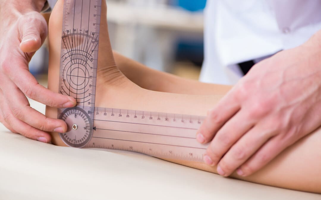
Those experiencing neck pain, stiffness, headache, shoulder and back pain may suffer from a whiplash injury. Can knowing whiplash signs and symptoms help individuals recognize the injury and help healthcare providers develop an effective treatment plan?
Contents
Whiplash Signs and Symptoms
Whiplash is a neck injury that typically occurs after a motor vehicle collision or accident but can happen with any injury that rapidly whips the neck forward and backward. It is a mild to moderate injury of the neck muscles. Common whiplash signs and symptoms include:
Some individuals can develop chronic pain and headaches.
The symptoms and treatment depend on the severity of the injury. Treatment can include over-the-counter pain medicines, ice and heat therapy, chiropractic, physical therapy, and stretching exercises.
Frequent Signs and Symptoms
The sudden whipping movement of the head can affect several structures within the neck. These structures include:
Muscles
Bones
Joints
Tendons
Ligaments
Intervertebral discs
Blood vessels
Nerves.
Any or all of these can be affected by a whiplash injury. (MedlinePlus, 2017)
Statistics
Whiplash is a neck sprain that occurs from a fast neck-jerking motion. Whiplash injuries account for more than half of vehicle traffic collision injuries. (Michele Sterling, 2014) Even with a minor injury, the most frequent symptoms include: (Nobuhiro Tanaka et al., 2018)
Neck pain
Next stiffness
Neck tenderness
Limited range of motion of the neck
Individuals can develop neck discomfort and pain shortly after an injury; however, the more intense pain and stiffness typically do not occur right after the injury. Symptoms tend to worsen the next day or 24 hours later. (Nobuhiro Tanaka et al., 2018)
Beginning Symptoms
Researchers have found that approximately more than half of individuals with whiplash develop symptoms within six hours of the injury. Around 90% develop symptoms within 24 hours, and 100% develop symptoms within 72 hours. (Nobuhiro Tanaka et al., 2018)
Whiplash vs. Traumatic Cervical Spine Injury
Whiplash describes a mild to moderate neck injury without significant skeletal or neurological symptoms. Significant neck injuries can lead to fractures and dislocations of the spine that can affect the nerves and spinal cord. Once an individual develops neurological problems associated with a neck injury, the diagnosis changes from whiplash to traumatic cervical spine injury. These differences can be confusing as they are on the same spectrum. To better understand the severity of a neck sprain, the Quebec classification system divides neck injury into the following grades (Nobuhiro Tanaka et al., 2018)
Grade 0
This means there are no neck symptoms or physical examination signs.
Grade 1
There is neck pain and stiffness.
Very few findings from the physical examination.
Grade 2
Indicates neck pain and stiffness
Neck tenderness
Decreased mobility or neck range of motion on physical examination.
Grade 3
Involves muscle pain and stiffness.
Neurologic symptoms include:
Numbness
Tingling
Weakness in the arms
Decreased reflexes
Grade 4
Involves a fracture or dislocation of the bones of the spinal column.
Other Symptoms
Other whiplash signs and symptoms that can be associated with the injury but are less common or only occur with a severe injury include (Nobuhiro Tanaka et al., 2018)
Tension headache
Jaw pain
Sleep problems
Migraine headache
Difficulty concentrating
Reading difficulties
Blurred vision
Dizziness
Driving difficulties
Rare Symptoms
Individuals with severe injuries can develop rare symptoms that often indicate traumatic cervical spine injury and include: (Nobuhiro Tanaka et al., 2018)
Amnesia
Tremor
Voice changes
Torticollis – painful muscle spasms that keep the head turned to one side.
Bleeding in the brain
Complications
Most individual generally recover from their symptoms within a few weeks to a few months. (Michele Sterling, 2014) However, whiplash complications can occur, especially with severe grade 3 or grade 4 injuries. The most common complications of a whiplash injury include chronic/long-term pain and headaches. (Michele Sterling, 2014) Traumatic cervical spine injury can affect the spinal cord and be associated with chronic neurological problems, including numbness, weakness, and difficulty walking. (Luc van Den Hauwe et al., 2020)
Treatment
The pain is typically more severe the next day than after the injury. Whiplash musculoskeletal injury treatment depends on whether it is an acute injury or the individual has developed chronic neck pain and stiffness.
Acute pain can be treated with over-the-counter medicines like Tylenol and Advil, which effectively treat the pain.
Advil is a nonsteroidal anti-inflammatory that can be taken with the pain reliever Tylenol, which works in different ways.
The mainstay of treatment is encouraging regular activity with stretching and exercise. (Michele Sterling, 2014)
Physical therapy uses various range of motion exercises to strengthen the neck muscles and relieve the pain.
Chiropractic adjustments and non-surgical decompression can help realign and nourish the spine.
Acupuncture can cause the body to release natural hormones that provide pain relief, help relax the soft tissues, increase circulation, and reduce inflammation. The cervical spine can return to alignment when the soft tissues are no longer inflamed and spasming. (Tae-Woong Moon et al., 2014)
Sterling M. (2014). Physiotherapy management of whiplash-associated disorders (WAD). Journal of physiotherapy, 60(1), 5–12. doi.org/10.1016/j.jphys.2013.12.004
Tanaka, N., Atesok, K., Nakanishi, K., Kamei, N., Nakamae, T., Kotaka, S., & Adachi, N. (2018). Pathology and Treatment of Traumatic Cervical Spine Syndrome: Whiplash Injury. Advances in orthopedics, 2018, 4765050. doi.org/10.1155/2018/4765050
van Den Hauwe L, Sundgren PC, Flanders AE. (2020). Spinal Trauma and Spinal Cord Injury (SCI). In: Hodler J, Kubik-Huch RA, von Schulthess GK, editors. Diseases of the Brain, Head and Neck, Spine 2020–2023: Diagnostic Imaging [Internet]. Cham (CH): Springer; 2020. Chapter 19. Available from: www.ncbi.nlm.nih.gov/books/NBK554330/ doi: 10.1007/978-3-030-38490-6_19
Moon, T. W., Posadzki, P., Choi, T. Y., Park, T. Y., Kim, H. J., Lee, M. S., & Ernst, E. (2014). Acupuncture for treating whiplash associated disorder: a systematic review of randomised clinical trials. Evidence-based complementary and alternative medicine : eCAM, 2014, 870271. doi.org/10.1155/2014/870271
Can incorporating nopal or prickly pear cactus into one’s diet help individuals trying to lower blood glucose, inflammation, and risk factors associated with heart and metabolic diseases?
Contents
Nopal
Nopal, also known as prickly pear cactus, is a versatile vegetable that can be added to nutrition plans to increase fiber intake, vitamins, minerals, and plant-based compounds. It grows in the U.S. Southwest, Latin America, and the Mediterranean. The pads, or the nopales or cactus paddles, have a texture like okra and slight tartness. The prickly pear cactus fruit, referred to as tuna in Spanish, is also consumed. (University of Arizona Cooperative Extension, 2019) It is often used in fruit salsas, salads, and desserts and is available as a supplement in tablet and powder form.
Nopal is highly nutritious, low in calories, free of fat, sodium, or cholesterol, and full of fiber, vitamins, minerals, and betalains. (Parisa Rahimi et al., 2019) Betalains are pigments with anti-inflammatory properties. The variety of fibers creates a low glycemic index (measures how much a specific food raises blood sugar levels after consumption) of about 32, a recommended addition to a diabetes-friendly diet. (Patricia López-Romero et al., 2014)
Compounds
Nopal contains a variety of beneficial carbohydrates, vitamins, and minerals.
Nopal has soluble and insoluble fiber, which benefits blood sugar.
It also contains vitamin A, carotenoids, vitamin C, calcium, and plant-based compounds like phenols and betalains. (Karina Corona-Cervantes et al., 2022)
Blood Sugar Regulation
Research has evaluated regular nopal consumption and supplementation for blood sugar control. A study on blood sugar evaluated adding nopal to a high-carbohydrate breakfast or a breakfast high in soy protein in Mexican individuals with type 2 diabetes. The study found that consuming nopales, about 300 grams or 1.75 to 2 cups before a meal, could reduce after-meal/postprandial blood sugars. (Patricia López-Romero et al., 2014) An older study had similar results. (Montserrat Bacardi-Gascon et al., 2007) Individuals were randomly assigned to consume 85 grams of nopal with three different breakfast options:
Chilaquiles – a casserole made with corn tortilla, vegetable oil, and pinto beans.
Burritos – made with eggs, vegetable oil, and pinto beans.
Quesadillas – made with flour tortillas, low-fat cheese, avocado, and pinto beans.
The groups assigned to eat nopales had reductions in blood sugar. There was a:
30% reduction in the chilaquiles group.
20% decrease in the burrito group.
48% reduction in the quesadilla group.
However, the studies were small, and the population was not diverse. so further research is needed.
Increased Fiber
The combination of soluble and insoluble fiber benefits the gut in various ways. Soluble fiber can act as a prebiotic, feeding beneficial bacteria in the gut and assisting in removing low-density lipoprotein (LDL) cholesterol from the body. Insoluble fiber increases transit time, or how quickly food moves through the digestive system and promotes bowel regularity. (Centers for Disease Control and Prevention, 2022) In a short-term randomized clinical control trial, researchers found an improvement in irritable bowel syndrome symptoms in individuals supplemented with 20 and 30 grams of nopal fiber. (Jose M Remes-Troche et al., 2021) For individuals not used to consuming fibrous foods, it may cause mild diarrhea, so it is recommended to increase intake slowly and with adequate water to prevent gas and bloating.
Plant Based Calcium
One cup of nopal provides 244 milligrams or 24% of daily calcium needs. Calcium is a mineral that optimizes bone and teeth health. It also assists in blood vessel contraction and dilation, muscle function, blood clotting, nerve transmission, and hormonal secretion. (National Institutes of Health. Office of Dietary Supplements 2024) Individuals who follow diets that exclude dairy products can benefit from plant-based calcium sources. This includes cruciferous vegetables like kale, collards, and arugula.
Other Benefits
Studies done in animals and test tubes suggest that fresh nopal and extracts may assist in reducing triglycerides and cholesterol in metabolic dysfunction-associated steatotic liver disease or when unhealthy amounts of fat accumulate in the liver. (Karym El-Mostafa et al., 2014) Other potential benefits with limited evidence include:
Unless individuals are allergic to it, most can eat whole nopal without a problem. However, supplementing is different because it provides a concentrated source. Individuals taking medication to manage diabetes and consuming nopal regularly may contribute to an increased risk of developing hypoglycemia or low blood sugar. Dermatitis has also been reported from contact with the cactus spines. (U.S. Department of Agriculture, FoodData Central, 2018) There have been rare reports of bowel obstruction in individuals who consume large amounts of the seeds found in the fruit. (Karym El-Mostafa et al., 2014) Ask a registered dietitian or primary healthcare provider if nopal can provide safe benefits.
Rahimi, P., Abedimanesh, S., Mesbah-Namin, S. A., & Ostadrahimi, A. (2019). Betalains, the nature-inspired pigments, in health and diseases. Critical reviews in food science and nutrition, 59(18), 2949–2978. doi.org/10.1080/10408398.2018.1479830
López-Romero, P., Pichardo-Ontiveros, E., Avila-Nava, A., Vázquez-Manjarrez, N., Tovar, A. R., Pedraza-Chaverri, J., & Torres, N. (2014). The effect of nopal (Opuntia ficus indica) on postprandial blood glucose, incretins, and antioxidant activity in Mexican patients with type 2 diabetes after consumption of two different composition breakfasts. Journal of the Academy of Nutrition and Dietetics, 114(11), 1811–1818. doi.org/10.1016/j.jand.2014.06.352
Corona-Cervantes, K., Parra-Carriedo, A., Hernández-Quiroz, F., Martínez-Castro, N., Vélez-Ixta, J. M., Guajardo-López, D., García-Mena, J., & Hernández-Guerrero, C. (2022). Physical and Dietary Intervention with Opuntia ficus-indica (Nopal) in Women with Obesity Improves Health Condition through Gut Microbiota Adjustment. Nutrients, 14(5), 1008. doi.org/10.3390/nu14051008
Bacardi-Gascon, M., Dueñas-Mena, D., & Jimenez-Cruz, A. (2007). Lowering effect on postprandial glycemic response of nopales added to Mexican breakfasts. Diabetes care, 30(5), 1264–1265. doi.org/10.2337/dc06-2506
Remes-Troche, J. M., Taboada-Liceaga, H., Gill, S., Amieva-Balmori, M., Rossi, M., Hernández-Ramírez, G., García-Mazcorro, J. F., & Whelan, K. (2021). Nopal fiber (Opuntia ficus-indica) improves symptoms in irritable bowel syndrome in the short term: a randomized controlled trial. Neurogastroenterology and motility, 33(2), e13986. doi.org/10.1111/nmo.13986
El-Mostafa, K., El Kharrassi, Y., Badreddine, A., Andreoletti, P., Vamecq, J., El Kebbaj, M. S., Latruffe, N., Lizard, G., Nasser, B., & Cherkaoui-Malki, M. (2014). Nopal cactus (Opuntia ficus-indica) as a source of bioactive compounds for nutrition, health and disease. Molecules (Basel, Switzerland), 19(9), 14879–14901. doi.org/10.3390/molecules190914879
Onakpoya, I. J., O’Sullivan, J., & Heneghan, C. J. (2015). The effect of cactus pear (Opuntia ficus-indica) on body weight and cardiovascular risk factors: a systematic review and meta-analysis of randomized clinical trials. Nutrition (Burbank, Los Angeles County, Calif.), 31(5), 640–646. doi.org/10.1016/j.nut.2014.11.015
Corona-Cervantes, K., Parra-Carriedo, A., Hernández-Quiroz, F., Martínez-Castro, N., Vélez-Ixta, J. M., Guajardo-López, D., García-Mena, J., & Hernández-Guerrero, C. (2022). Physical and Dietary Intervention with Opuntia ficus-indica (Nopal) in Women with Obesity Improves Health Condition through Gut Microbiota Adjustment. Nutrients, 14(5), 1008. doi.org/10.3390/nu14051008
Can improving breathing patterns help further fitness and optimize overall health for individuals who walk for exercise?
Contents
Improve Breathing and Walking
Exercising is a moment in which breathing can quicken and become labored if not done correctly. There is a proper way to breathe when exercising, especially when walking or speed walking. Breathing incorrectly causes rapid fatigue and exhaustion. Controlling the flow of one’s breath improves endurance and cardiovascular health, and it can also amplify metabolism, mood, and energy levels. (Hsiu-Chin Teng et al., 2018) Known as diaphragmatic breathing, it is used for those with reduced lung capacity, like individuals with chronic obstructive pulmonary disease/COPD. The practice improves lung capacity and is a recommended way to help relieve stress.
Physiology
During exercise, the oxygen inhaled converts the calories consumed into energy that fuels the body. This process is referred to as metabolism.
When the oxygen supply exceeds the body’s oxygen needs, the body is in an aerobic state. This means there is plenty of oxygen to fuel physical activity/exercise as there are calories to burn.
If the oxygen supply falls short of the body’s oxygen needs, the body falls into an anaerobic state.
Deprived of oxygen, the body turns to stored fuel in the muscles, known as glycogen.
This delivers a powerful burst of energy, but the fuel is quickly spent and fatigue and exhaustion soon follow.
Increasing airflow in and out of the lungs can prevent early exhaustion and help the body burn calories more effectively. (Your lungs and exercise. Breathe 2016)
Improved Breathing Benefits
Optimal breathing starts in infancy. When a baby breathes, their belly rises and falls. This facilitates respiration by pushing and pulling the diaphragm – the muscle that separates the lungs and abdominal cavity. When the baby inhales, the belly extends, pulling the diaphragm downward and allowing the lungs to fill with air. When the baby exhales, the belly draws in, pressing the diaphragm upward and forcing air out. As the body ages and the capacity of the lungs increases, individuals shift from belly-breathing to chest-breathing. Chest breathing involves the chest wall muscles with little use of the diaphragm. Chest breathing usually provides enough air for everyday activity but does not fill the lungs.
This is why individuals resort to mouth-breathing or gasping when the oxygen supply is limited. Even those in decent physical shape may be inadvertently undermining efforts by sucking in their stomach to look thinner, depriving themselves of complete inhalations and exhalations. To overcome this, individuals need to re-train their bodies to activate the abdominal muscles when walking. Belly or diaphragmatic breathing can extend the duration of exercise while strengthening the core muscles. (Nelson, Nicole 2012) By increasing core stability, individuals can better support the spine and maintain a healthy posture when walking. This stabilizes the hips, knees, upper back, and shoulders, making the body less prone to strain, instability, and fatigue from unhealthy posture. (Tomas K. Tong et al., 2014)
Breathing Correctly
The inhalation draws the belly out, pulls the diaphragm down, and inflates the lungs. Simultaneously, it extends the ribcage and lengthens the lower spine. This forces the shoulders and collarbone backward, further opening the chest. Exhaling does the reverse.
Walking
Start by inhaling and exhaling through the nose, ensuring that the inhalation duration matches the exhalation duration. When picking up the pace, individuals can resort to mouth-breathing, maintaining the same inhalation/exhalation rhythm. At no time should breathing be held in. Learning diaphragmatic breathing takes time, but the following steps can be a starting point:
Inhale by inflating the belly fully on a count of five.
Allow the lungs to fill, drawing the shoulders back as this happens.
Exhale by pulling the belly button toward the spine on a count of five.
Use the diaphragm to press the air out of the lungs, keeping the spine erect.
Repeat.
If unable to maintain a count of five, individuals can shorten the count or slow the pace of the walk. Individuals in good shape may be able to extend the count. Initially, diaphragmatic breathing may not come naturally, but it will become automatic with practice. Stop and place the hands over the head if short of breath when walking. Breathe in and out deeply and evenly until breathing returns to normal.
Unlocking Wellness
References
Teng, H. C., Yeh, M. L., & Wang, M. H. (2018). Walking with controlled breathing improves exercise tolerance, anxiety, and quality of life in heart failure patients: A randomized controlled trial. European journal of cardiovascular nursing, 17(8), 717–727. doi.org/10.1177/1474515118778453
Tong, T. K., Wu, S., Nie, J., Baker, J. S., & Lin, H. (2014). The occurrence of core muscle fatigue during high-intensity running exercise and its limitation to performance: the role of respiratory work. Journal of sports science & medicine, 13(2), 244–251.
Nelson, Nicole MS, LMT. (2012). Diaphragmatic Breathing: The Foundation of Core Stability. Strength and Conditioning Journal 34(5):p 34-40, October 2012. | DOI: 10.1519/SSC.0b013e31826ddc07
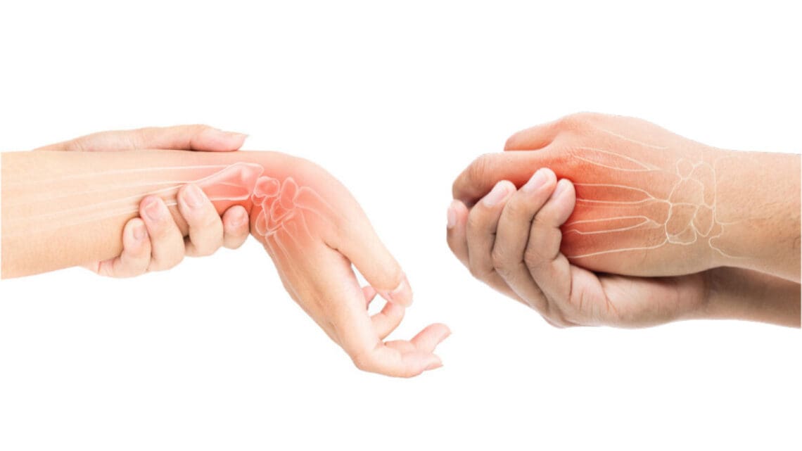
Can individuals with joint hypermobility find relief through nonsurgical treatments in reducing pain and restoring body mobility?
Contents
Introduction
When a person moves their body, the surrounding muscles, joints, and ligaments are incorporated into various tasks that allow them to stretch and be flexible without pain or discomfort. Many repetitive motions enable the individual to continue their routine. However, when the joints, muscles, and ligaments are stretched farther than normal in the upper and lower extremities without pain, it is known as joint hypermobility. This connective tissue disorder can correlate with other symptoms that affect the body and cause many people to seek treatment to manage joint hypermobility symptoms. In today’s article, we will look at joint hypermobility and how various non-surgical treatments can help reduce pain caused by joint hypermobility and restore body mobility. We talk with certified medical providers who consolidate our patients’ information to assess how their pain may be associated with joint hypermobility. We also inform and guide patients on how integrating various non-surgical treatments can help improve joint function while managing the associated symptoms. We encourage our patients to ask their associated medical providers intricate and insightful questions about incorporating non-surgical therapies as part of their routine to reduce pain and discomfort from joint hypermobility. Dr. Jimenez, D.C., includes this information as an academic service. Disclaimer.
What Is Joint Hypermobility?
Do you often feel your joints locked up in your hands, wrists, knees, and elbows? Do you experience pain and fatigue in your joints when your body feels constantly tired? Or when you stretch your extremities, do they extend farther than usual to feel the relief? Many of these various scenarios are often correlated with individuals experiencing joint hypermobility. Joint hypermobility is an inherited disorder with autosomal dominant patterns that characterize joint hyperlaxity and musculoskeletal pain within the body extremities. (Carbonell-Bobadilla et al., 2020) This connective tissue condition is often related to the flexibility of the connected tissues like ligaments and tendons in the body. An example would be if a person’s thumb is touching their inner forearm without feeling pain or discomfort, they have joint hypermobility. Additionally, many individuals dealing with joint hypermobility will often have a difficult diagnosis as they will develop skin and tissue fragility over time, causing musculoskeletal complications. (Tofts et al., 2023)
When individuals deal with joint hypermobility over time, many often have symptomatic joint hypermobility. They will present with musculoskeletal and systemic symptoms that lead to displaying skeletal deformities, tissue and skin fragility, and structural differences in the body’s system. (Nicholson et al., 2022) Some of the symptoms that joint hypermobility are shown in a diagnosis include:
Muscle pain and joint stiffness
Clicking joints
Fatigue
Digestive issues
Balance issues
Luckily, there are various treatments that many people can use to help restrengthen the surrounding muscles around the joints and reduce the correlating symptoms caused by joint hypermobility.
Movement As Medicine-Video
Nonsurgical Treatments For Joint Hypermobility
When dealing with joint hypermobility, many individuals need to seek treatments to reduce the correlating pain-like symptoms of joint hypermobility and help relieve the body’s extremities while restoring mobility. Some excellent treatments for joint hypermobility are non-surgical therapies that are non-invasive, gentle on the joints and muscles, and cost-effective. Various non-surgical treatments can be customized for the individual depending on how severe their joint hypermobility and comorbidities affect the person’s body. Non-surgical treatments can relieve the body from joint hypermobility by treating the causes of the pain through reduction and maximizing functional capacity and restoring a person’s quality of life. (Atwell et al., 2021) The three non-surgical treatments that are excellent for reducing pain from joint hypermobility and helping strengthen the surrounding muscles are below.
Chiropractic Care
Chiropractic care utilizes spinal manipulation and helps restore joint mobility in the body to reduce the effects of joint hypermobility by stabilizing the affected joints from the hypermobile extremities. (Boudreau et al., 2020) Chiropractors incorporate mechanical and manual manipulation and various techniques to help many individuals improve their posture by being more mindful of their bodies and work with multiple other therapies to emphasize controlled movements. With other comorbidities associated with joint hypermobility, like back and neck pain, chiropractic care can reduce these comorbidity symptoms and allow the individual to regain their quality of life.
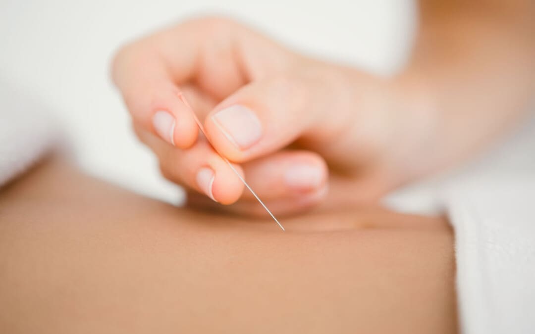
Acupuncture
Another non-surgical treatment that many individuals can incorporate to reduce joint hypermobility and its comorbidities is acupuncture. Acupuncture utilizes small, thin, solid needles that acupuncturists use to block pain receptors and restore the body’s energy flow. When many individuals are dealing with joint hypermobility, their extremities in the legs, hands, and feet are in pain over time, which can cause the body to be unstable. What acupuncture does is help reduce the pain caused by joint hypermobility associated with the extremities and restore balance and functionality to the body (Luan et al., 2023). This means that if a person is dealing with stiffness and muscle pain from joint hypermobility, acupuncture can help rewire the pain by placing the needles in the body’s acupoints to provide relief.
Physical Therapy
Physical therapy is the last non-surgical treatment many people can incorporate into their daily routine. Physical therapy can help manage joint hypermobility that are tailored to help strengthen weak muscles that are surrounding the affected joints, improving a person’s stability and helping reduce the risk of dislocation. Additionally, many individuals can use low-impact exercise to ensure optimal motor control when doing regular exercises without putting excessive strain on the joints. (Russek et al., 2022)
By incorporating these three non-surgical treatments as part of a customized treatment for joint hypermobility, many individuals will begin to feel a difference in their balance. They will not experience joint pain by being more mindful of the body and incorporating small changes in their routine. Even though living with joint hypermobility can be a challenge for many individuals, by integrating and utilizing the right combination of non-surgical treatments, many can begin to lead active and fulfilling lives.
References
Atwell, K., Michael, W., Dubey, J., James, S., Martonffy, A., Anderson, S., Rudin, N., & Schrager, S. (2021). Diagnosis and Management of Hypermobility Spectrum Disorders in Primary Care. J Am Board Fam Med, 34(4), 838-848. doi.org/10.3122/jabfm.2021.04.200374
Boudreau, P. A., Steiman, I., & Mior, S. (2020). Clinical management of benign joint hypermobility syndrome: a case series. J Can Chiropr Assoc, 64(1), 43-54. www.ncbi.nlm.nih.gov/pubmed/32476667
Carbonell-Bobadilla, N., Rodriguez-Alvarez, A. A., Rojas-Garcia, G., Barragan-Garfias, J. A., Orrantia-Vertiz, M., & Rodriguez-Romo, R. (2020). [Joint hypermobility syndrome]. Acta Ortop Mex, 34(6), 441-449. www.ncbi.nlm.nih.gov/pubmed/34020527 (Sindrome de hipermovilidad articular.)
Luan, L., Zhu, M., Adams, R., Witchalls, J., Pranata, A., & Han, J. (2023). Effects of acupuncture or similar needling therapy on pain, proprioception, balance, and self-reported function in individuals with chronic ankle instability: A systematic review and meta-analysis. Complement Ther Med, 77, 102983. doi.org/10.1016/j.ctim.2023.102983
Nicholson, L. L., Simmonds, J., Pacey, V., De Wandele, I., Rombaut, L., Williams, C. M., & Chan, C. (2022). International Perspectives on Joint Hypermobility: A Synthesis of Current Science to Guide Clinical and Research Directions. J Clin Rheumatol, 28(6), 314-320. doi.org/10.1097/RHU.0000000000001864
Russek, L. N., Block, N. P., Byrne, E., Chalela, S., Chan, C., Comerford, M., Frost, N., Hennessey, S., McCarthy, A., Nicholson, L. L., Parry, J., Simmonds, J., Stott, P. J., Thomas, L., Treleaven, J., Wagner, W., & Hakim, A. (2022). Presentation and physical therapy management of upper cervical instability in patients with symptomatic generalized joint hypermobility: International expert consensus recommendations. Front Med (Lausanne), 9, 1072764. doi.org/10.3389/fmed.2022.1072764
Tofts, L. J., Simmonds, J., Schwartz, S. B., Richheimer, R. M., O’Connor, C., Elias, E., Engelbert, R., Cleary, K., Tinkle, B. T., Kline, A. D., Hakim, A. J., van Rossum, M. A. J., & Pacey, V. (2023). Pediatric joint hypermobility: a diagnostic framework and narrative review. Orphanet J Rare Dis, 18(1), 104. doi.org/10.1186/s13023-023-02717-2
For athletes and sports enthusiasts, a torn triceps can be a serious injury. Can knowing their symptoms, causes, risk factors, and potential complications help healthcare providers develop an effective treatment plan?
Contents
Torn Triceps Injury
The triceps is the muscle on the back of the upper arm that allows the elbow to straighten. Fortunately, triceps tears are uncommon, but they can be serious. The injury affects men more often than women and usually occurs from trauma, sports, and/or exercise activities. Depending on the extent and severity of the injury, a torn triceps injury can require splinting, physical therapy, and possibly surgery to regain movement and strength. Recovery after a triceps tear typically lasts around six months. (The Ohio State University Wexner Medical Center. 2021)
Anatomy
The triceps brachii muscle, or triceps, runs along the back of the upper arm. It is named tri- because it has three heads – the long, medial, and lateral head. (Sendic G. 2023) The triceps originates at the shoulder and attaches to the shoulder blade/scapula and upper arm bone/humerus. At the bottom, it attaches to the point of the elbow. This is the bone on the pinky side of the forearm, known as the ulna. The triceps cause movement at the shoulder and the elbow joint. At the shoulder, it performs extension or backward movement of the arm and adduction or moving the arm toward the body. The main function of this muscle is at the elbow, where it performs extension or straightening of the elbow. The triceps work the opposite of the biceps muscle on the front of the upper arm, which conducts flexion or bending of the elbow.
Triceps Tear
Tears can occur anywhere along the length of a muscle or tendon, which is the structure that attaches the muscle to the bones. Triceps tears commonly occur in the tendon connecting the triceps to the back of the elbow. Muscle and tendon tears are graded from 1 to 3 based on severity. (Alberto Grassi et al., 2016)
Grade 1 Mild
These small tears cause pain that worsens with movement.
There is some swelling, bruising, and minimal loss of function.
Grade 2 Moderate
These tears are larger and have moderate swelling and bruising.
The fibers are partially torn and stretched.
Up to 50% loss of function.
Grade 3 Severe
This is the worst type of tear, where the muscle or tendon is completely torn.
These injuries cause severe pain and disability.
Symptoms
Triceps tears cause immediate pain in the back of the elbow and upper arm that worsens when trying to move the elbow. Individuals might also feel and/or hear a popping or tearing sensation. There will be swelling, and the skin will likely be red and/or bruised. With a partial tear, the arm will feel weak. If there is a complete tear, there will be significant weakness when straightening the elbow. Individuals may also notice a lump on the back of their arm where the muscles have contracted and knotted together.
Causes
Triceps tears usually occur during trauma, when the muscle is contracted and an external force pushes the elbow into a bent position. (Kyle Casadei et al., 2020) One of the most common causes is by falling on an outstretched arm. Triceps tears also occur during sports activities like:
Throwing a baseball
Blocking in a football game
Gymnastics
Boxing
When a player falls and lands on their arm.
Tears can also happen when using heavy weights during triceps-targeted exercises, such as the bench press.
Tears can also occur from direct trauma to the muscle, like a motor vehicle accident, but are less common.
Long-Term
Triceps tears can develop over time as a result of tendonitis. This condition usually occurs from repetitive use of the triceps muscle during activities like manual labor or exercise. Triceps tendonitis is sometimes referred to as weightlifter’s elbow. (Orthopedic & Spine Center. N.D.) The strain on tendons causes tiny tears that the body typically heals. However, if more strain is placed on the tendon than it can keep up with, the tiny tears can begin to grow.
Risk Factors
Risk factors can increase the risk of a triceps tear. Underlying medical conditions can weaken tendons, increasing the risk of injury, and can include: (Tony Mangano et al., 2015)
Diabetes
Rheumatoid arthritis
Hyperparathyroidism
Lupus
Xanthoma – fatty deposits of cholesterol under the skin.
Hemangioendothelioma – cancerous or noncancerous tumors caused by abnormal growth of blood vessel cells.
Chronic kidney failure
Chronic tendonitis or bursitis in the elbow.
Individuals who have had cortisone shots in the tendon.
Individuals using anabolic steroids.
Triceps tears tend to occur more commonly in males between 30 and 50. (Ortho Bullets. 2022) This comes from participating in activities like football, weightlifting, bodybuilding, and manual labor, which also increases the risk of injury.
Treatment
Treatment depends on which part of the triceps is affected and the extent of the damage. It may only need resting for a few weeks, physical therapy, or require surgery.
Nonsurgical
Partial tears in the triceps that involve less than 50% of the tendon can often be treated without surgery. (Mehmet Demirhan, Ali Ersen 2016) Initial treatment includes:
Splinting the elbow with a slight bend for four to six weeks allows the injured tissue to heal. (Ortho Bullets. 2022)
During this time, ice can be applied to the area for 15 to 20 minutes several times daily to help decrease pain and swelling.
Non-steroidal anti-inflammatory medications/NSAIDs – Aleve, Advil, and Bayer can help reduce inflammation.
Other over-the-counter medications like Tylenol can help decrease the pain.
Once the splint is removed, physical therapy will help restore movement and strength in the elbow.
Full movement is expected to return within 12 weeks, but full strength will not return until six to nine months after the injury. (Mehmet Demirhan, Ali Ersen 2016)
Surgery
Triceps tendon tears that involve more than 50% of the tendon require surgery. In some cases, however, surgery may still be recommended for tears smaller than 50% if the individual has a physically demanding job or plans to resume playing sports at a high level. Tears in the muscle belly or area where the muscle and tendon join are typically sewn back together. If the tendon is no longer attached to the bone, it is screwed back on. Recovery and physical therapy after surgery depend on the specific surgeon’s protocols. In general, individuals will spend a couple of weeks in a brace. Around four weeks after surgery, individuals will be able to start moving the elbow again. However, they won’t be able to start doing heavy lifting for four to six months. (Ortho Bullets. 2022) (Mehmet Demirhan, Ali Ersen 2016)
Complications
Complications can occur after triceps repair, whether there was surgery or not. For example, individuals may have problems regaining full elbow extension or straightening. They are also at a higher risk of re-rupture if they try to use the arm before it’s fully healed. (Mehmet Demirhan, Ali Ersen 2016)
Grassi, A., Quaglia, A., Canata, G. L., & Zaffagnini, S. (2016). An update on the grading of muscle injuries: a narrative review from clinical to comprehensive systems. Joints, 4(1), 39–46. doi.org/10.11138/jts/2016.4.1.039
Casadei, K., Kiel, J., & Freidl, M. (2020). Triceps Tendon Injuries. Current sports medicine reports, 19(9), 367–372. doi.org/10.1249/JSR.0000000000000749
Mangano, T., Cerruti, P., Repetto, I., Trentini, R., Giovale, M., & Franchin, F. (2015). Chronic Tendonopathy as a Unique Cause of Non Traumatic Triceps Tendon Rupture in a (Risk Factors Free) Bodybuilder: A Case Report. Journal of orthopaedic case reports, 5(1), 58–61. doi.org/10.13107/jocr.2250-0685.257
For individuals living with cyclical or chronic endometriosis symptoms, can incorporating support therapies help in disease management?
Contents
Support Therapies
Endometriosis is a disorder in which tissue similar to the uterine lining begins to grow outside the uterus where it does not belong. Endometriosis support therapies involve a comprehensive approach to treatment. It involves non-invasive treatments to help manage symptoms that can include:
A physical therapist uses various pressures, stretching, and/or trigger point release. This helps: (Sylvia Mechsner, 2022)
Release muscle tension
Lower cortisol – stress hormone
Improve circulation
Release endorphins – the body’s natural painkillers
Medications
Nonsteroidal anti-inflammatory drugs or NSAIDs and hormonal contraceptives – birth control are the first line of treatment. Advil and Motrin are over-the-counter NSAIDs. If those don’t manage pain effectively, a healthcare provider may recommend prescription NSAIDs. (Sylvia Mechsner, 2022) Hormonal suppression agents or estrogen modulators are a second line of treatment for endometriosis and can include: (Christian M. Becker et al., 2022)
Hormonal contraceptives suppress or regulate periods. They are effective for management, but not everyone can take them because of medical history, side effects, or fertility disorders and conditions. (Mert Ilhan et al., 2019) A healthcare provider can recommend alternative support therapies.
Transcutaneous Electrical Nerve Stimulation
A transcutaneous electrical nerve stimulation utilizes a battery-operated device that delivers low-voltage electrical stimulation to nerve fibers through electrodes placed on the skin.
Sessions are usually 15 to 30 minutes and work by disrupting pain signals. (Sylvia Mechsner, 2022)
Acupuncture
Acupuncture is a therapy in which a practitioner inserts thin needles into specific acupoints on the body to promote the flow of energy and alleviate pain. (Nora Giese et al., 2023)
Chiropractic
Chiropractic care focuses on spinal adjustments and alignment to enhance nervous system function, help alleviate pelvic discomfort and nerve pain – sciatica – and improve overall well-being. (Robert J. Trager et al., 2021)
Non-surgical decompression could be recommended to gently stretch the spine, relieve pressure, and flood the spine with added nutrients.
Movement Medicine: Chiropractic Care
References
Mansfield, C., Lenobel, D., McCracken, K., Hewitt, G., & Appiah, L. C. (2022). Impact of Pelvic Floor Physical Therapy on Function in Adolescents and Young Adults with Biopsy-Confirmed Endometriosis at a Tertiary Children’s Hospital: A Case Series. Journal of pediatric and adolescent gynecology, 35(6), 722–727. doi.org/10.1016/j.jpag.2022.07.004
Mechsner S. (2022). Endometriosis, an Ongoing Pain-Step-by-Step Treatment. Journal of clinical medicine, 11(2), 467. doi.org/10.3390/jcm11020467
Ilhan, M., Gürağaç Dereli, F. T., & Akkol, E. K. (2019). Novel Drug Targets with Traditional Herbal Medicines for Overcoming Endometriosis. Current drug delivery, 16(5), 386–399. doi.org/10.2174/1567201816666181227112421
Becker, C. M., Bokor, A., Heikinheimo, O., Horne, A., Jansen, F., Kiesel, L., King, K., Kvaskoff, M., Nap, A., Petersen, K., Saridogan, E., Tomassetti, C., van Hanegem, N., Vulliemoz, N., Vermeulen, N., & ESHRE Endometriosis Guideline Group (2022). ESHRE guideline: endometriosis. Human reproduction open, 2022(2), hoac009. doi.org/10.1093/hropen/hoac009
Pereira, A., Herrero-Trujillano, M., Vaquero, G., Fuentes, L., Gonzalez, S., Mendiola, A., & Perez-Medina, T. (2022). Clinical Management of Chronic Pelvic Pain in Endometriosis Unresponsive to Conventional Therapy. Journal of personalized medicine, 12(1), 101. doi.org/10.3390/jpm12010101
Giese, N., Kwon, K. K., & Armour, M. (2023). Acupuncture for endometriosis: A systematic review and meta-analysis. Integrative medicine research, 12(4), 101003. doi.org/10.1016/j.imr.2023.101003
Trager, R.J., Prosak, S.E., Leonard, K.A. et al. (2021). Diagnosis and management of sciatic endometriosis at the greater sciatic foramen: a case report. SN Comprehensive Clinical Medicine, 3. doi.org/doi:10.1007/s42399-021-00941-0
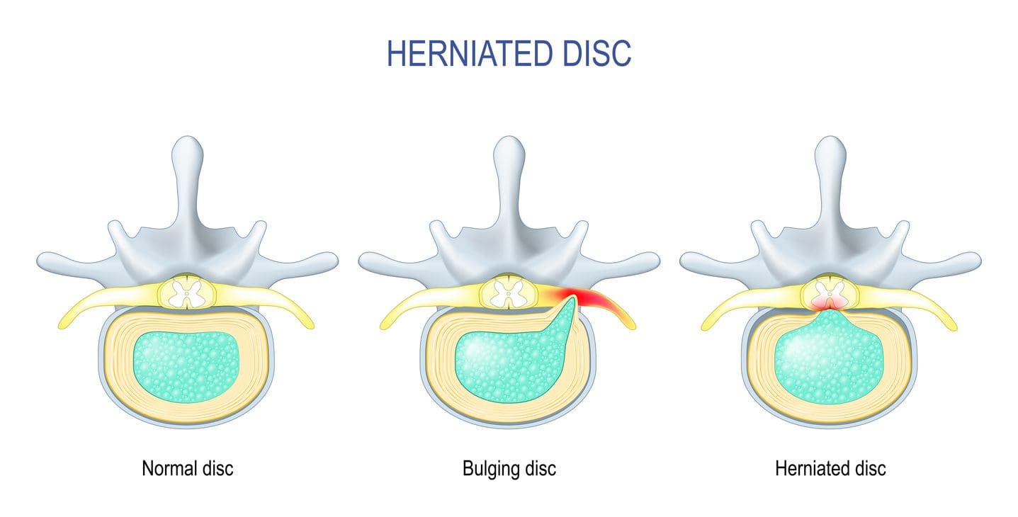
Can individuals with herniated discs find the relief they are looking for from traction therapy or decompression to provide pain relief?
Contents
Introduction
The spine allows the individual to be mobile and flexible without feeling pain and discomfort when a person is on the move. This is because the spine is part of the musculoskeletal system that consists of muscles, tendons, ligaments, the spinal cord, and spinal discs. These components surround the spine and have three regions to allow the upper and lower extremities to do their jobs. However, the spine also ages when the body starts to age naturally. Many movements or routine actions can cause the body to be stiff and, over time, can cause the spinal disc to herniate. When this happens, a herniated disc can lead to pain and discomfort in the extremities, thus making individuals deal with a reduced quality of life and pain in three spinal regions. Luckily, there are numerous treatments, like traction therapy and decompression, to alleviate the pain and discomfort associated with herniated discs. Today’s article looks at why herniated discs cause issues in the spine and the effects of how these two treatments can help reduce herniated discs. We talk with certified medical providers who consolidate our patients’ information to assess how a herniated disc in the spine may be the issue causing musculoskeletal pain. We also inform and guide patients on how integrating spinal decompression and traction therapy can help realign the spine and reduce disc herniation that is causing spinal issues. We encourage our patients to ask their associated medical providers intricate and important questions about incorporating non-surgical treatments as part of their routine to reduce pain and discomfort in their bodies. Dr. Jimenez, D.C., includes this information as an academic service. Disclaimer.
Why Herniated Discs Causes Issues In The Spine?
Have you been experiencing constant discomfort in your neck or back that doesn’t allow you to relax? Do you feel tingling sensations in your upper and lower extremities, making grasping objects or walking difficult? Or have you noticed that you are hunching over from your desk or standing and that stretching causes pain? As the spine keeps the body upright, its main components include the moveable vertebrae, the nerve root fibers, and spinal discs to help send neuron signals to the brain to allow movement, cushion the shocked forces on the spine, and be flexible. The spine allows the individual to perform various tasks without pain and discomfort through repetitive movements. However, when the body ages, it can lead to degenerative changes in the spine, causing the spinal disc to herniate over time. A herniated disc is a common degenerative musculoskeletal condition that causes the nucleus pulposus to break through any weak region of the annulus fibrosus and compress the surrounding nerve roots. (Ge et al., 2019) Other times, when repetitive motions start to cause a developing herniated disc, the inner portion of the disc can become desiccated and brittle. In contrast, the outer portion becomes more fibrotic and less elastic, causing the disc to shrink and be narrow. A herniated disc can affect young and old populations as they can have a multifactorial contribution that causes proinflammatory changes to the body. (Wu et al., 2020)
When many people are dealing with pain associated with a herniated disc, the disc itself goes through morphological change through the characterization of the disc being partial damage, which is then followed by the displacement and herniation of the inner disc portion in the vertebral canal to compress the spinal nerve roots. (Diaconu et al., 2021) This causes symptoms of pain, numbness, and weakness in the upper and lower body portions through nerve impingement. Hence why, many individuals are dealing with referred pain symptoms from their arms and legs that are radiating pain. When nerve compression associated with herniated discs starts to cause pain and discomfort, many individuals begin to seek out treatment to reduce the pain that the herniated disc is causing to provide relief for their bodies.
Spinal Decompression In Depth-Video
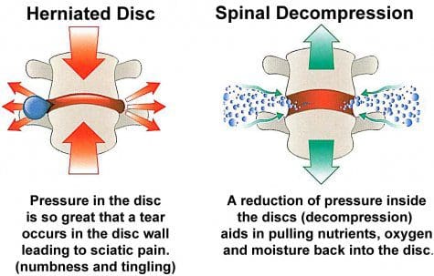
The Effects Of Traction Therapy In Reducing Herniated Disc
Many people who are suffering from pain that is being affected by herniated discs in their spines can seek out treatments like traction therapy to alleviate pain. Traction therapy is a non-surgical treatment that stretches and mobilizes the spine. Traction therapy can be mechanically or manually done by a pain specialist or with the help of mechanical devices. The effects of traction therapy can reduce the compression force on the spinal disc while reducing nerve root compression by expanding the disc height within the spine. (Wang et al., 2022) This allows the surrounding joints within the spine to be mobile and positively affect the spine. With traction therapy, intermittent or steady tension forces help stretch the spine, reduce pain, and improve functional outcomes. (Kuligowski et al., 2021)
The Effects Of Spinal Decompression In Reducing Herniated Disc
Another form of non-surgical treatment is spinal decompression, a sophisticated version of traction that uses computerized technology to help apply controlled, gentle pulling forces to the spine. Spinal decompression does is that it can help decompress the spinal canal and help pull the herniated disc back to its original position while stabilizing the spine and keeping the vital bones and soft tissues safe. (Zhang et al., 2022) Additionally, spinal decompression can create negative pressure on the spine to allow the flow of nutritional fluids and blood oxygen back to the discs while creating an inverse relationship when tension pressure is introduced. (Ramos & Martin, 1994) Both spinal decompression and traction therapy can offer many therapeutic pathways to provide relief to many individuals dealing with herniated discs. Depending on how severe the herniated disc has caused issues to the person’s spine, many can rely on non-surgical treatments due to its customizable plan that is personalized to the person’s pain and can be combined with other therapies to strengthen the surrounding muscles. By doing so, many people can be pain-free over time while being mindful of their bodies.
References
Diaconu, G. S., Mihalache, C. G., Popescu, G., Man, G. M., Rusu, R. G., Toader, C., Ciucurel, C., Stocheci, C. M., Mitroi, G., & Georgescu, L. I. (2021). Clinical and pathological considerations in lumbar herniated disc associated with inflammatory lesions. Rom J Morphol Embryol, 62(4), 951-960. doi.org/10.47162/RJME.62.4.07
Ge, C. Y., Hao, D. J., Yan, L., Shan, L. Q., Zhao, Q. P., He, B. R., & Hui, H. (2019). Intradural Lumbar Disc Herniation: A Case Report and Literature Review. Clin Interv Aging, 14, 2295-2299. doi.org/10.2147/CIA.S228717
Kuligowski, T., Skrzek, A., & Cieslik, B. (2021). Manual Therapy in Cervical and Lumbar Radiculopathy: A Systematic Review of the Literature. Int J Environ Res Public Health, 18(11). doi.org/10.3390/ijerph18116176
Ramos, G., & Martin, W. (1994). Effects of vertebral axial decompression on intradiscal pressure. J Neurosurg, 81(3), 350-353. doi.org/10.3171/jns.1994.81.3.0350
Wang, W., Long, F., Wu, X., Li, S., & Lin, J. (2022). Clinical Efficacy of Mechanical Traction as Physical Therapy for Lumbar Disc Herniation: A Meta-Analysis. Comput Math Methods Med, 2022, 5670303. doi.org/10.1155/2022/5670303
Wu, P. H., Kim, H. S., & Jang, I. T. (2020). Intervertebral Disc Diseases PART 2: A Review of the Current Diagnostic and Treatment Strategies for Intervertebral Disc Disease. Int J Mol Sci, 21(6). doi.org/10.3390/ijms21062135
Zhang, Y., Wei, F. L., Liu, Z. X., Zhou, C. P., Du, M. R., Quan, J., & Wang, Y. P. (2022). Comparison of posterior decompression techniques and conventional laminectomy for lumbar spinal stenosis. Front Surg, 9, 997973. doi.org/10.3389/fsurg.2022.997973
Can using egg substitutes or replacements be safe for individuals with an egg allergy?
Contents
Substitutes and Replacements
Individuals should not assume either is safe unless they carefully read the label.
Egg substitutes may contain eggs.
Egg replacement products may be egg-free.
Look for alternatives labeled vegan or egg-free to ensure there are none.
Substitutes May Contain Eggs
Liquid egg substitutes in grocery store dairy aisles are made from eggs. The following all contain eggs and are not safe for individuals with egg allergies:
Generic liquid egg substitutes in cartons
Egg Beaters
Powdered egg white products
Replacements Are Safe Alternatives
Special replacement products that do not contain eggs are available.
They are labeled vegan egg substitutes.
They are usually sold in powdered form.
They are useful for baking.
They cannot be used as a replacement for eggs in foods like a quiche.
Always check the ingredients on the label before purchasing a product sold as a substitute or replacement to ensure it is completely free.
These products may also contain soy, dairy, or other food allergens.
Vegan – contains no animal products, which includes eggs and dairy.
Vegetarian – may contain eggs as they are not meat but an animal product.
Unaware of Foods With Eggs
Stay aware of eggs hidden in other food products, such as cakes, breads, pastries, noodles, crackers, and cereals.
The federal Food Allergen Labeling and Consumer Protection Act requires that all packaged food products that contain eggs as an ingredient must list the word egg on the label. (U.S. Food & Drug Administration. 2022)
Other ingredients that indicate eggs are in the product include:
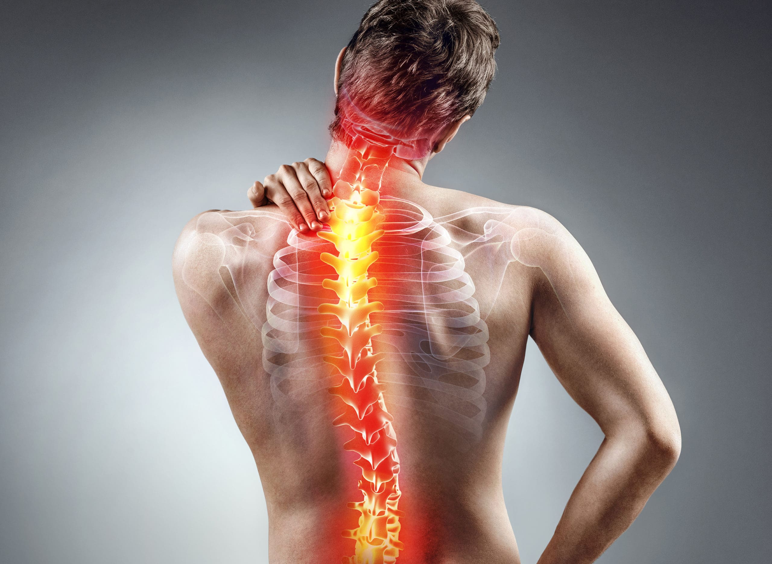
Can individuals with spinal pain in their necks and back utilize decompression therapy to restore spinal disc height and find relief?
Contents
Introduction
Many people don’t realize that as the body gets older, so does the spine. The spine is part of the musculoskeletal system that provides structural support to the body by keeping it upright. The surrounding muscles, ligaments, and tissues surrounding the spine help with stability and mobility, while the spinal disc and joints provide shock absorption from the sheer vertical weight. When a person is on the move with their daily activities, the spine can allow the individual to be mobile without pain or discomfort. However, as time passes, the spine goes through degenerative changes that can cause pain and discomfort to the body, thus leaving the individual to deal with overlapping risk profiles that can affect their neck and back. To that point, many people seek out treatments to reduce the pain affecting their spine and restore the disc height in their bodies. Today’s article looks at how spinal pain affects a person’s neck and back and how treatments like spinal decompression can reduce spinal pain and restore disc height. We talk with certified medical providers who consolidate our patients’ information to assess how spinal pain can significantly impact a person’s well-being and quality of life in their bodies. We also inform and guide patients on how integrating spinal decompression can help reduce spinal pain and restore spinal disc height. We encourage our patients to ask their associated medical providers intricate and important questions about incorporating non-surgical treatments into a health and wellness routine to relieve spinal pain and regain their quality of life. Dr. Jimenez, D.C., includes this information as an academic service. Disclaimer.
How Spinal Pain Affects A Person’s Neck & Back
Do you feel constant muscle aches and pains in your neck and back? Have you experienced stiffness and limited mobility when you are twisting and turning? Or do heavy objects cause muscle strain when moving from one location to another? Many individuals will be on the move and be in weird positions without feeling pain and discomfort when it comes to the spine. This is due to the surrounding muscles and tissues being stretched and the spinal discs taking on the vertical pressure on the spine. However, when environmental factors, traumatic injuries, or natural aging start to affect the spine, it can lead to the development of spinal pain. This is because the outer portion of the spinal disc is intact, and the inner portion of the disc is being affected. When abnormal stresses start to reduce the water intake within the disc, it can internally stimulate the pain receptors without nerve root symptoms inside the disc. (Zhang et al., 2009) This causes many individuals to deal with neck and back pain in their bodies and reduces their quality of life.
Spinal pain can lead to overlapping risk profiles that cause many individuals to deal with severe low back pain and neck pain, which then causes the surrounding muscles to become weak, tight, and overstretched. At the same time, the surrounding nerve roots are also affected as the nerve fibers surround the outer and inner parts of the spinal disc, which causes nociceptive pain properties to the neck and back region and leads to discogenic pain. (Coppes et al., 1997) When many individuals are dealing with muscle pain correlated with the spinal discs, it causes a pain-spasm-pain cycle that can affect their bodies due to not moving enough and causing painful muscular activities when trying to be mobile. (Roland, 1986) When a person has limited mobility cause they are experiencing spinal pain, their natural disc height slowly degenerates, causing more issues to their bodies and socioeconomic burdens. Fortunately, when many individuals are dealing with spinal pain, numerous treatments can reduce spinal pain and restore their disc height.
Movement Medicine- Video
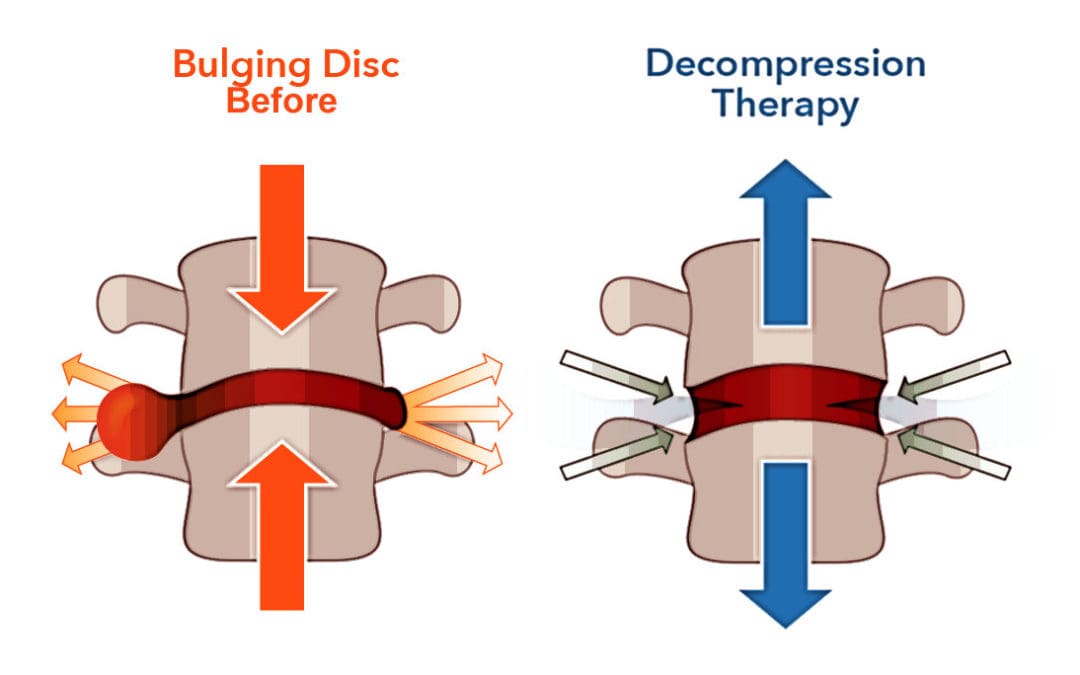
How Spinal Decompression Reduces Spinal Pain
When people are seeking treatments for their spinal pain, many will seek surgical treatments to reduce their pain, but it will be a bit pricey. However, many individuals will opt for non-surgical treatments due to their affordability. Non-surgical treatments are cost-effective and customizable to a person’s pain and discomfort. From chiropractic care to acupuncture, depending on the severity of the person’s pain, many will find the relief they seek. One of the most innovative treatments for reducing spinal pain is spinal decompression. Spinal decompression allows the individual to be strapped into a traction table. This is because it gently pulls on the spine to realign the spinal disc by reducing the pressure on the spine to invoke the body’s natural healing process to relieve pain. (Ramos & Martin, 1994) Additionally, when many individuals are using spinal decompression, the gentle traction provides a motorized distraction to the spine that may induce physical changes to the spinal disc and help restore a person’s range of motion, flexibility, and mobility. (Amjad et al., 2022)
Spinal Decompression Restoring Spinal Disc Height
When a person is being strapped into the spinal decompression machine, the gentle traction helps the spinal disc return to the spine, allowing the fluids and nutrients to rehydrate the spine, increasing the spine’s disc height. This is because spinal decompression creates negative pressure on the spine, allowing the spinal disc to return to its original height and providing relief. Plus, the amazing thing that spinal decompression does is that it can be combined with physical therapy to help stretch and strengthen the surrounding muscles near the spine to provide more stability and flexibility. (Vanti et al., 2023) This allows the individual to be more mindful of their bodies and start incorporating small habit changes to reduce the pain from returning. When many people begin to think about their health and wellness by going to treatment, they will regain their quality of life and get back to their daily routine without the issues affecting their spine.
References
Amjad, F., Mohseni-Bandpei, M. A., Gilani, S. A., Ahmad, A., & Hanif, A. (2022). Effects of non-surgical decompression therapy in addition to routine physical therapy on pain, range of motion, endurance, functional disability and quality of life versus routine physical therapy alone in patients with lumbar radiculopathy; a randomized controlled trial. BMC Musculoskelet Disord, 23(1), 255. doi.org/10.1186/s12891-022-05196-x
Coppes, M. H., Marani, E., Thomeer, R. T., & Groen, G. J. (1997). Innervation of “painful” lumbar discs. Spine (Phila Pa 1976), 22(20), 2342-2349; discussion 2349-2350. doi.org/10.1097/00007632-199710150-00005
Ramos, G., & Martin, W. (1994). Effects of vertebral axial decompression on intradiscal pressure. J Neurosurg, 81(3), 350-353. doi.org/10.3171/jns.1994.81.3.0350
Roland, M. O. (1986). A critical review of the evidence for a pain-spasm-pain cycle in spinal disorders. Clin Biomech (Bristol, Avon), 1(2), 102-109. doi.org/10.1016/0268-0033(86)90085-9
Vanti, C., Saccardo, K., Panizzolo, A., Turone, L., Guccione, A. A., & Pillastrini, P. (2023). The effects of the addition of mechanical traction to physical therapy on low back pain? A systematic review with meta-analysis. Acta Orthop Traumatol Turc, 57(1), 3-16. doi.org/10.5152/j.aott.2023.21323
Zhang, Y. G., Guo, T. M., Guo, X., & Wu, S. X. (2009). Clinical diagnosis for discogenic low back pain. Int J Biol Sci, 5(7), 647-658. doi.org/10.7150/ijbs.5.647
IFM's Find A Practitioner tool is the largest referral network in Functional Medicine, created to help patients locate Functional Medicine practitioners anywhere in the world. IFM Certified Practitioners are listed first in the search results, given their extensive education in Functional Medicine