Back Clinic Chiropractic Examination. An initial chiropractic examination for musculoskeletal disorders will typically have four parts: a consultation, case history, and physical examination. Laboratory analysis and X-ray examination may be performed. Our office provides additional Functional and Integrative Wellness Assessments in order to bring greater insight into a patient’s physiological presentations.
Consultation:
The patient will meet the chiropractor which will assess and question a brief synopsis of his or her lower back pain, such as:
Duration and frequency of symptoms
Description of the symptoms (e.g. burning, throbbing)
Areas of pain
What makes the pain feel better (e.g. sitting, stretching)
What makes the pain feel worse (e.g. standing, lifting).
Case history. The chiropractor identifies the area(s) of complaint and the nature of the back pain by asking questions and learning more about different areas of the patient’s history, including:
Family history
Dietary habits
Past history of other treatments (chiropractic, osteopathic, medical and other)
Occupational history
Psychosocial history
Other areas to probe, often based on responses to the above questions.
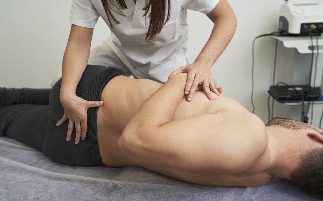
Physical examination: We will utilize a variety of methods to determine the spinal segments that require chiropractic treatments, including but not limited to static and motion palpation techniques determining spinal segments that are hypo mobile (restricted in their movement) or fixated. Depending on the results of the above examination, a chiropractor may use additional diagnostic tests, such as:
X-ray to locate subluxations (the altered position of the vertebra)
A device that detects the temperature of the skin in the paraspinal region to identify spinal areas with a significant temperature variance that requires manipulation.
Laboratory Diagnostics: If needed we also use a variety of lab diagnostic protocols in order to determine a complete clinical picture of the patient. We have teamed up with the top labs in the city in order to give our patients the optimal clinical picture and appropriate treatments.
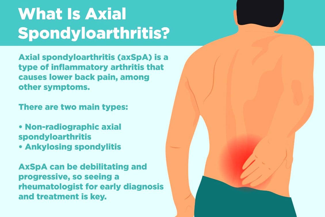
Non-radiographic axial spondyloarthritis or nr-axSpA and non-radiographic ankylosing spondylitis/AS are related. However, non-radiographic axial spondyloarthritis can present AS symptoms with active inflammation of the spine and sacroiliac/SI joints, causing back and hip pain but does not reveal joint damage on X-rays or MRIs. Injury Medical Chiropractic and Functional Medicine Clinic can explain what it means to have non-radiographic axial spondyloarthritis, how it can be managed, and what to do to prevent it from turning into ankylosing spondylitis.
Non-Radiographic Axial Spondyloarthritis
Non-radiographic axial spondyloarthritis means there are early AS symptoms but have not developed enough joint inflammation or damage to show up on an X-ray or other form of imaging. Early evidence of joint inflammation includes blurring of the joint edges and localized regions of joint erosion. It can be difficult for physicians to see these subtle changes on an x-ray.
Ankylosing Spondylitis
Ankylosing spondylitis, or AS, is a form of inflammatory arthritis that affects joints in the spine and elsewhere.
It is a chronic, inflammatory, autoimmune disease.
Medical research is still ongoing to determine the exact cause, but a genetic component is believed to be contributing factor.
Around 85% of individuals with ankylosing spondylitis have inherited the HLA-B27 gene, which is associated with multiple autoimmune conditions.
In the early stages, individuals will present lower back pain around the sacroiliac joints or the joints that connect the spine to the pelvis.
Later stages have more obvious X-ray findings, like the fusing of the sacroiliac joints and the lower spine that takes place over time.
Joint inflammation can progress, causing permanent joint damage and spine rigidity.
Most individuals with the condition can manage their symptoms with NSAIDs, chiropractic care, physical and massage therapy, and range of motion exercises.
Stage 1
There is no evidence of spinal inflammation on x-rays.
MRI provides more detailed images of bones and may reveal bone marrow edema or accumulation of fluid in the structures of the spinal bones and joints.
Individuals with non-radiographic axial spondyloarthritis, you are here.
Stage 2
There is visible inflammation of the spinal joints on the x-ray.
The sacroiliac joints between the spine and the pelvis are the most affected.
Stage 3
Chronic inflammation of the joints has caused bone loss and permanent joint damage, resulting in spine rigidity.
Symptoms of Non-Radiographic Axial Spondyloarthritis
There are differences between back pain associated with muscle strain and arthritis. Back pain symptoms include:
Starts to present before age 40.
It has a gradual onset and can go unnoticed for years.
Improves with movement or activity.
Eases up throughout the day.
Starts up in the evening when resting.
Other symptoms include:
Joint stiffness
Swollen fingers
Heel pain
Bilateral buttock discomfort and pain
Slowing Progression
Progression from non-radiographic axial spondyloarthritis to ankylosing spondylitis occurs in 10% – 20% of individuals over a two-year period. Progression factors include genetics, gender, degree of joint damage, and level of inflammatory markers at the time of diagnosis.
Early diagnosis and treatment can slow the progression before significant joint damage with anti-inflammatory therapy, rheumatological therapy, and targeted exercise.
Work with a specialist like an orthopedic spine specialist and rheumatologist that understands the disorder and is up to date on the most recent treatment modalities.
D. J. Pradeep, A. Keat, K. Gaffney, Predicting outcome in ankylosing spondylitis, Rheumatology, Volume 47, Issue 7, July 2008, Pages 942–945, doi.org/10.1093/rheumatology/ken195
Kucybała, Iwona, et al. “Radiologic approach to axial spondyloarthritis: where are we now and where are we heading?.” Rheumatology international vol. 38,10 (2018): 1753-1762. doi:10.1007/s00296-018-4130-1
Michelena, Xabier, López-Medina, Clementina, and Helena Marzo-Ortega. “Non-radiographic versus radiographic axSpA: what’s in a name?”.” National Center for Biotechnology Information. October 14, 2020. doi.org/10.1093/rheumatology/keaa422
Back discomfort sensations and symptoms could indicate pulled-back muscles. Unless you’ve experienced the condition before, determining the cause can be difficult. A pulled-back muscle can start as a sudden, sharp sting when bending, reaching, or twisting. Or it can present gradually, worsening over a few days. It is a common injury, but if left untreated could take several weeks, and in severe cases, a few months, to heal correctly. Injury Medical Chiropractic and Functional Medicine Clinic can help diagnose the problem and develop a customized treatment plan to restore optimal function and health.
Pulled Back Muscles
A pulled muscle describes a strained muscle.
A strain is a muscle or tendon injury that happens when the tissue overstretches or tears.
When a ligament stretches or tears, it’s called a sprain.
Most cases can be managed and treated at home.
But if the symptoms are not improving or make it difficult to move, see a doctor or chiropractor.
Symptoms
Common signs and symptoms of a pulled-back muscle include:
Swelling
Tenderness
Soreness – Sore muscles that feel tight and achy usually indicate a condition that is likely to improve in a few days. More severe soreness could indicate a more significant injury.
Spasms – A sudden convulsive spasm in the muscle can also indicate a pull. This can feel like a sudden tightening that does not release. The muscle can continue to spasm and lead to other symptoms.
Cramping – A muscle can cramp can lead to increased tightness whenever trying to use the muscle.
Pain – Can be characterized as a constant dullness and/or soreness in most situations or, in severe cases, sharp and shooting.
Discomfort when moving around. If pain flares up when trying to move or use the back muscles is usually an indication that something is wrong.
Relief during inactivity and rest. When lying down to rest or taking a temporary break, and the symptoms disappear could also be an indication of a pulled-back muscle or another injury.
Causes
The most common causes are:
A Strained Muscle
This causes some damage to a region of muscle tissue, usually the result of being over-used or torn from another injury.
Sprained Ligaments
Involves damage to the ligaments in a joint, usually those in the spinal vertebrae.
A Herniated Disc
This involves damage to the discs that can leak out, irritating the surrounding tissues and nerves and can cause shifting and misalignment of the spine.
These conditions are distinct, but the symptoms can be similar.
Therapies
It is important to consult a medical professional before treating an injury because symptoms of other injuries, such as disc problems or a broken bone, can resemble strains and sprains. Most treatments will utilize:
Ice and Heat
Ice helps reduce inflammation.
The faster ice can be applied to a pulled-back muscle, the quicker pain and swelling are reduced, and the healing process can begin.
Apply a cold pack for 15-20 minutes as soon as the injury occurs.
Take a 20-minute break between each cold application.
After the first days, alternate cold therapy with heat to increase circulation.
Try a 20-20-20 rule: 20 minutes of an ice pack followed by a 20-minute break, then 20 minutes of heat.
Repeat as necessary, allowing 20 minutes between heat or ice therapy.
Limited Rest
Right after a muscle strain, limiting physical activity levels and avoiding movements are recommended for a short period.
After the initial pain subsides, partial activity levels are recommended to help prevent the muscles from weakening.
Compression
Applying compression bandages or using an active compression system can help reduce swelling and edema and repair damaged tissues faster.
Stretching
Returning to activities, gentle stretching exercises can improve tissue healing by increasing blood circulation to the injured area.
Applying heat to the area before stretching can also help.
Strength Training
Ask a doctor or chiropractor about the right strength exercises for your condition.
Strength training will develop the muscles to prevent future injuries.
Pain Medication
Pain levels are an important indicator during the recovery process.
Pain medications relieve symptoms but do not assist with healing and should only be used short-term to provide relief.
If you need pain medication, consult your physician to determine the appropriate type and dosage for your situation.
Massage
Blood circulation to the injured tissues is increased with massage therapy.
Chiropractic
A chiropractor can diagnose back pain from a muscle or disc injury and develop an individualized treatment plan.
Allen, Laura. “Case Study: The Use of Massage Therapy to Relieve Chronic Low-Back Pain.” International journal of therapeutic massage & bodywork vol. 9,3 27-30. 9 Sep. 2016, doi:10.3822/ijtmb.v9i3.267
Kumar, Saravana et al. “The effectiveness of massage therapy for the treatment of nonspecific low back pain: a systematic review of systematic reviews.” International journal of general medicine vol. 6 733-41. 4 Sep. 2013, doi:10.2147/IJGM.S50243
Dr. Alex Jimenez, D.C., presents how the SBAR method is used in a clinical approach in a chiropractic office. Since pain in the body is one of the most common complaints worldwide, many individuals can be referred to the right healthcare professional to have a better understanding of what is happening to their bodies and have their health and wellness restored. We refer patients to certified providers specializing in treatments to aid individuals suffering from various chronic issues associated with muscle and joint pain affecting their bodies. We also guide our patients by referring them to our associated medical providers based on their examination when it’s appropriate. We find that education is the solution to asking our providers insightful questions. Dr. Alex Jimenez, D.C., provides this information as an educational service only. Disclaimer
What Is The SBAR Method?
Dr. Alex Jimenez, D.C., presents: The term SBAR stands for situation, background, assessment, and recommendation. It is a communication method that many chiropractors or healthcare professionals use to help simplify communicating patient information to other healthcare team members. And the whole goal of the SBAR method is to help us strategically and systematically share a patient situation along with the background of that patient, the assessment findings that we have found, and recommendations that we recommend to that specific individual so they can easily understand what we need, want, and what is going on with that patient in a very clear and focused way. So the SBAR method can help the chiropractor or massage therapist stay organized whenever they’re having to communicate and cut out unnecessary information that may be in the conversation that wastes time or may confuse the listener and help prevent those moments where the specialist may get questions from the person they are talking to, and they may not know.
The SBAR method allows chiropractors to communicate efficiently with patients about where the pain is located in their bodies. So the SBAR will help many health professionals stay organized. Some examples of the SBAR method used for communication include: a nurse needs to speak with a healthcare provider like a physician, a nurse practitioner, or a PA to let them know that the patient’s condition is deteriorating, and they need to call and report that. If they need something for that patient, the healthcare provider can follow the SBAR method, which will help them clearly and concisely communicate that issue to the listener. Chiropractors can also use the SBAR to share with other associated medical providers or massage therapists when they have a patient’s report to be handed or transferred to a different unit.
The SBAR method can be used with other healthcare team members, like speech therapy, occupational therapy, chiropractic therapy, and physical therapy. This method helps and guides chiropractors with what information they need to provide to the patient, so they can fully understand what is going on with them. An example would be a patient coming into a chiropractic clinic with back pain; however, they are experiencing gut issues and having areas of complaints in their hips, causing mobility issues. So by using the SBAR method, chiropractors and other healthcare providers can communicate better with their patients and develop a solution with the APPIER process and a treatment plan that caters to the individual. When creating your SBAR to communicate better with someone, it’s better to ensure that you are fully prepared before initiating that conversation. Having a little system to comply with the SBAR method can help you quickly and allow you to note what is happening with the patient in your head or take note of their condition. Getting the layout of the SBAR method is the first step, and many healthcare units will have them created so the doctor can fill them in and put all the information they need when they call or talk to their patients.
Chiropractors using the SBAR method would go into the room, look at that patient, assess that patient, collect their vital signs and look in the chart, look at the latest progress now, and know who’s on board taking care of that patient. The SBAR method also allows the doctor to review that patient’s chart thoroughly and understand what’s going on with that patient. So by the time they step into the room, they will have an idea of what is going on with the patient when those questions come up. Plus, when they have looked at the latest lab results from their associated medical providers. They can have an insight into what medication the patient is taking because those questions will probably come up and be included in the SBAR method. This will allow the chiropractor to gather all that information from the patient and be comfortable and ready to initiate the conversation.
Situation
Dr. Alex Jimenez, D.C., presents: Now let’s look at each of the sections of the SBAR method. Since the SBAR method is very focused and concise with communication, it is straightforward. So the situation is the first thing you’re going to start with whenever you’re communicating using the SBAR method. So by having your computer on that specific patient, doctors can easily look at something in case the person asks them a question and have the information in front of them quickly. So with the situation, just as it says, the goal is to communicate why the patient is calling. That’s its purpose, as it helps start things off and allows the doctor and the patient to introduce themselves and briefly explain what is going on with their bodies. An example would be a person with back pain introducing themselves to the chiropractor and vice versa and briefly describing where they are in pain.
Background
Dr. Alex Jimenez, D.C., presents: The background portion of the SBAR method helps paint a picture of what the patient is going through and will provide a brief description of the situation. Then after that, we’ll go straight into the patient’s background, and this part of the communication will be very focused again. And how you would transition from situation to background in the SBAR method by going into the patient’s diagnosis. So the patient was admitted with whatever diagnosis on the date of admission. Then the chiropractor will tailor and include important patient information based on what the patient is experiencing pain-wise. The pain can vary from each person and can affect the body differently.
Many doctors can include the patient’s code status and discuss any other significant health problems that accompany the patient’s current situation. An example would be if a person is dealing with cardiac issues, their primary doctor can ask them if they had any health history with cardiovascular disorders, medications for heart diseases, chest pain, etcetera. Getting their background history can provide many doctors with a treatment plan that won’t cause any issues for the patient. When chiropractors work with other healthcare professionals, they can provide a background history of the patient, including bloodwork, previous procedures, and any additional information to develop a treatment plan. Along with consults, what other doctor groups are on board with this patient and any pending procedures the patient may have? That lets them know, okay, I don’t need to order this test or product because they will be having this procedure.
Assessment
Dr. Alex Jimenez, D.C., presents: The next section of the SBAR method is the assessment part, where the doctor will tell the patient what they have assessed or found in the patient. Many healthcare professionals, like chiropractors, provide those assessment findings and current vital signs to back up what they think is going on. An example would be a functional medicine doctor explaining to the patient what they found in their body, like possible respiratory, cardiac, or GI issues, and what they think is going on based on what they discovered.
But let’s say, for example, that the nurse or doctor doesn’t know; however, they know that something’s wrong with the patient and they need something. In this situation, the doctor or the nurse can take note of what is going on with the patient and explain to their associated medical providers that they are worried or that the patient is deteriorating; they’re unstable and have changed from when they previously saw them. By using the SBAR method, chiropractors can asses the situation the patient is dealing with and provide insightful solutions to develop a treatment plan for the patient.
Recommendation
Dr. Alex Jimenez, D.C., presents: And finally, the final part of the SBAR method is recommendations. So recommendations are where the doctor communicates with the patient on what they want or need. By laying out the framework from using the SBAR method, the recommendation part allows the doctor to specifically communicate with the patient on what needs to be done to improve their health and wellness. An example is if a patient is dealing with gut issues associated with metabolic syndrome and their doctor gives them a treatment plan to incorporate more nutritional foods in their diets, exercising more and getting an adjustment from a chiropractor can help alleviate pain affecting their backs or hips.
Conclusion
Since body pain is one of the most common complaints worldwide, chiropractic care can assist in managing the symptoms associated with joint and muscle pain while being cost-efficient and non-invasive. Utilizing the SBAR method in a chiropractic clinic can give the chiropractor the right tools to develop a treatment plan for the individual to relieve any pain affecting their body. Chiropractic care can also use the APPIER method combined with the SBAR method to fully alleviate any disorder in the body structure to restore a person’s health and wellness.
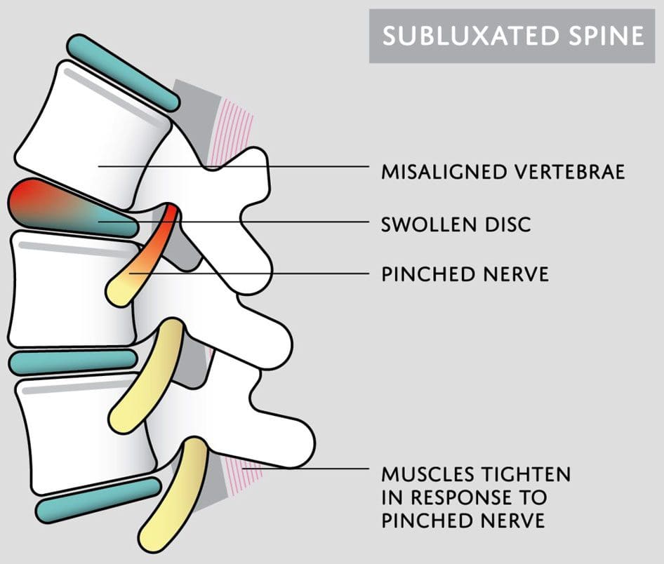
Subluxation is when a joint shifts out of alignment, which can happen to any joint in the body. Spinal subluxation indicates a misalignment of one or more portions of the spinal vertebrae. It is common in the spine from all the reaching, bending, twisting, and flexing the body goes through. Spinal subluxations, if left untreated, can cause disc degeneration, permanent nerve damage, neurological conditions, and chronic pain symptoms. A subluxation chiropractor will realign and decompress the spine combined with massage therapy to relax the muscles and restore mobility and function.
Subluxation Chiropractor
Some subluxations don’t cause any problems or pain, but that doesn’t mean they aren’t affecting the back and body. A spinal subluxation can cause long-term problems by:
Research shows that spinal subluxations can affect many facets of the body. Long-term effects may include:
Sleep problems
Low energy
Brain fog
Mood swings
Anxiety and depression
Digestive issues
Respiratory problems
Bone spurs
Spinal arthritis
Chiropractic Care
When the spine is out of alignment, it can cause issues throughout the body. Changes in one area affect the rest of the body. A subluxation chiropractor looks at the spine’s neurological and mechanical components and aims to reset everything back into its proper position. Similar to the way a massage helps the mind and body relax and de-stress, a spinal adjustment helps by:
Increasing circulation
Relieving discomfort and pain
Releasing tension
Improving mood
Reducing stress levels
Improving sleep function
Increasing energy levels
When the spine is properly aligned, the body can operate at its full potential.
Green, J D et al. “Anterior subluxation of the cervical spine: hyperflexion sprain.” AJNR. American journal of neuroradiology vol. 2,3 (1981): 243-50.
Meyer, S. “Thoracic spine trauma.” Seminars in roentgenology vol. 27,4 (1992): 254-61. doi:10.1016/0037-198x(92)90004-l
Neva MH, Häkkinen A, Mäkinen H, et al. High prevalence of asymptomatic cervical spine subluxation in patients with rheumatoid arthritis waiting for orthopedic surgeryAnnals of the Rheumatic Diseases 2006;65:884-888.
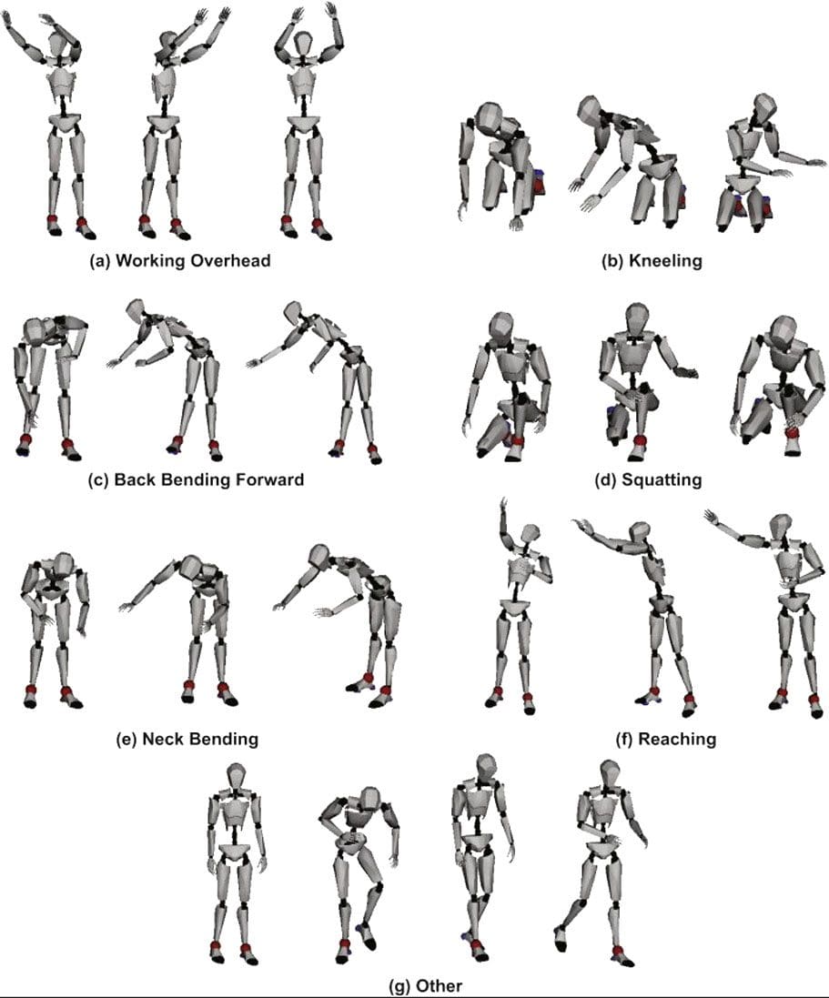
Nourollahi, Maryam, et al. “Awkward trunk postures and their relationship with low back pain in hospital nurses.” Work (Reading, Mass.) vol. 59,3 (2018): 317-323. doi:10.3233/WOR-182683
Vernon, Howard. “Historical overview and update on subluxation theories().” Journal of chiropractic humanities vol. 17,1 (2010): 22-32. doi:10.1016/j.echu.2010.07.001
Diagnosing ankylosing spondylitis usually involves multiple tests. When doctors order blood tests to diagnose ankylosing spondylitis, an individual is experiencing worsening symptoms in their back and joints. Often, a blood test diagnosis means the doctor is looking for evidence of anything else that could be causing the symptoms. However, blood tests by themselves cannot definitively diagnose ankylosing spondylitis, but when combined with imaging and assessment, they can provide important clues that point to the answers.
Ankylosing Spondylitis Blood Test Diagnosis
Ankylosing spondylitis is arthritis that primarily affects the spine and hips. It can be difficult to diagnose as no single test can provide thorough information for a definitive diagnosis. A combination of diagnostic tests are utilized, including a physical exam, imaging, and blood tests. Doctors are not only looking for results that will point to ankylosing spondylitis, but they are looking for any results that might point away from the spondylitis results that might provide a different explanation for symptoms.
Physical Exam
The diagnostic process will begin with the individual’s medical history, family history, and physical exam. During the exam, the doctor will ask questions to help rule out other conditions:
How long have symptoms been presenting?
Do symptoms get better with rest or exercise?
Are the symptoms getting worse or staying the same?
Are the symptoms worse at a particular time of day?
The doctor will check for limitations in mobility and palpate tender areas. Many conditions can cause similar symptoms, so the doctor will check to see if the pain or lack of mobility is consistent with ankylosing spondylitis. The feature sign of ankylosing spondylitis is pain and stiffness in the sacroiliac joints. The sacroiliac joints are located in the lower back, where the base of the spine and pelvis meet. The doctor will look at other spinal conditions and symptoms:
Back pain symptoms caused by – injuries, posture patterns, and/or sleeping positions.
The HLA-B27 gene corresponds with ankylosing spondylitis; if an individual has it, one of their parents has it.
Imaging
X-rays often serve as the first step to a diagnosis.
As the disease progresses, new small bones form between the vertebrae, eventually fusing them.
X-rays work best at mapping the disease progression than the initial diagnosis.
An MRI provides clearer images in the early stages as smaller details are visible.
Blood Tests
Blood tests can help rule out other conditions and check for signs of inflammation, providing supportive evidence along with the results of imaging tests. It typically only takes about a day or two to get the results. The doctor may order one of the following blood tests:
Antinuclear antibodies, or ANA, go after the proteins in the cell’s nucleus, telling the body its cells are the enemy.
This activates an immune response that the body fights to eliminate.
A study determined that ANA is found in 19% of individuals suffering from ankylosing spondylitis and is higher in women than men.
Combined with other tests, the presence of ANA provides another clue to a diagnosis.
Gut Health
The gut microbiome plays an important role in triggering the development of ankylosing spondylitis and its treatment.
Tests to determine the gut’s health can give a doctor a complete picture of what is happening inside the body.
Blood test diagnoses for ankylosing spondylitis and other inflammatory conditions rely heavily on piecing together different tests alongside clinical exams and imaging.
Causes, Symptoms, Diagnosis, and Treatment
References
Cardoneanu, Anca, et al. “Characteristics of the intestinal microbiome in ankylosing spondylitis.” Experimental and therapeutic medicine vol. 22,1 (2021): 676. doi:10.3892/etm.2021.10108
Prohaska, E et al. “Antinukleäre Antikörper bei Spondylitis ankylosans (Morbus Bechterew)” [Antinuclear antibodies in ankylosing spondylitis (author’s transl)]. Wiener klinische Wochenschrift vol. 92,24 (1980): 876-9.
Sheehan, Nicholas J. “The ramifications of HLA-B27.” Journal of the Royal Society of Medicine vol. 97,1 (2004): 10-4. doi:10.1177/014107680409700102
Wenker KJ, Quint JM. Ankylosing Spondylitis. [Updated 2022 Apr 9]. In: StatPearls [Internet]. Treasure Island (FL): StatPearls Publishing; 2022 Jan-. Available from: www.ncbi.nlm.nih.gov/books/NBK470173/
Xu, Yong-Yue, et al. “Role of the gut microbiome in ankylosing spondylitis: an analysis of studies in the literature.” Discovery medicine vol. 22,123 (2016): 361-370.
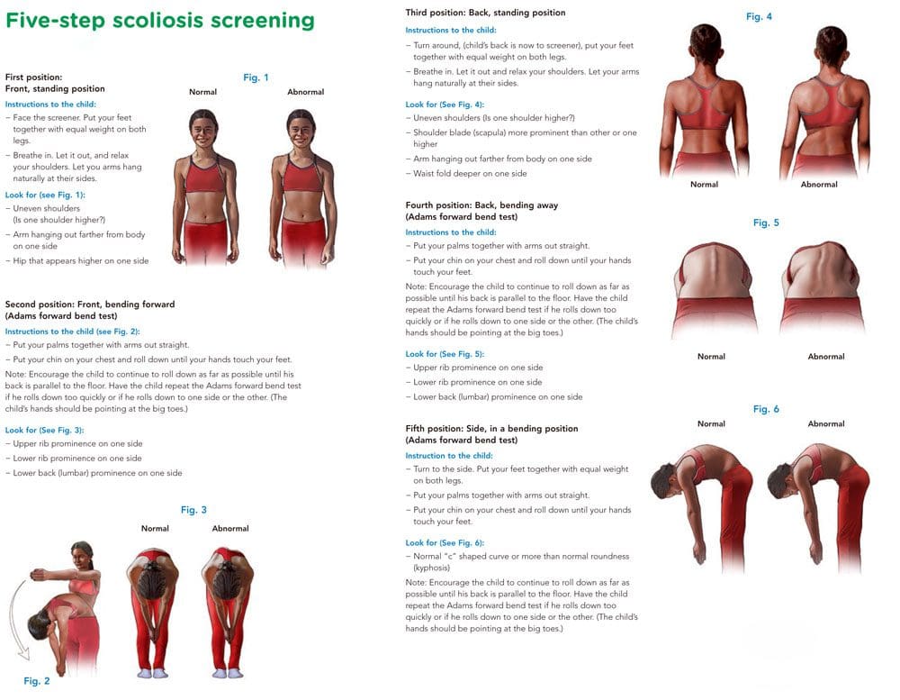
The Adams forward bend test is a simple screening method that can help with scoliosis diagnosis and help in developing a treatment plan. The exam is named after the English physician William Adams. As part of an examination, a doctor or chiropractor will look for an abnormal side-to-side bend in the spine.
Scoliosis Diagnosis
The Adams forward-bend test can help determine if there are indicators for scoliosis.
It is not an official diagnosis, but the results can be used as a starting point.
The Adams test will reveal signs of scoliosis and/or other potential deformities like:
Uneven shoulders
Uneven hips
Lack of symmetry between the vertebrae or the shoulder blades.
The head does not line up with a rib hump or the pelvis.
Detection of Other Spinal Issues
The test can also be used to find spinal curvature issues and conditions like:
Kyphosis or hunchback, where the upper back is bent forward.
Scheuermann’s disease is a form of kyphosis where the thoracic vertebrae can grow unevenly during a growth spurt and cause the vertebrae to develop into a wedge-like shape.
The Adams test by itself is not enough to confirm scoliosis.
A standing X-ray with Cobb angle measurements above 10 degrees is required for diagnosing scoliosis.
The Cobb angle determines which vertebrae are tilted the most.
The higher the angle, the more severe the condition and the more probable it will produce symptoms.
Computed tomography or CT and magnetic resonance imaging or MRI scans can also be used.
Forward Bend Test
References
Glavaš, Josipa et al. “The role of school medicine in the early detection and management of adolescent idiopathic scoliosis.” Wiener klinische Wochenschrift, 1–9. 4 Oct. 2022, doi:10.1007/s00508-022-02092-1
Grossman, T W et al. “An evaluation of the Adams forward bend test and the scoliometer in a scoliosis school screening setting.” Journal of pediatric orthopedics vol. 15,4 (1995): 535-8. doi:10.1097/01241398-199507000-00025
Letts, M et al. “Computerized ultrasonic digitization in the measurement of spinal curvature.” Spine vol. 13,10 (1988): 1106-10. doi:10.1097/00007632-198810000-00009
Senkoylu, Alpaslan, et al. “A simple method for assessing rotational flexibility in adolescent idiopathic scoliosis: modified Adam’s forward bending test.” Spine deformity vol. 9,2 (2021): 333-339. doi:10.1007/s43390-020-00221-2
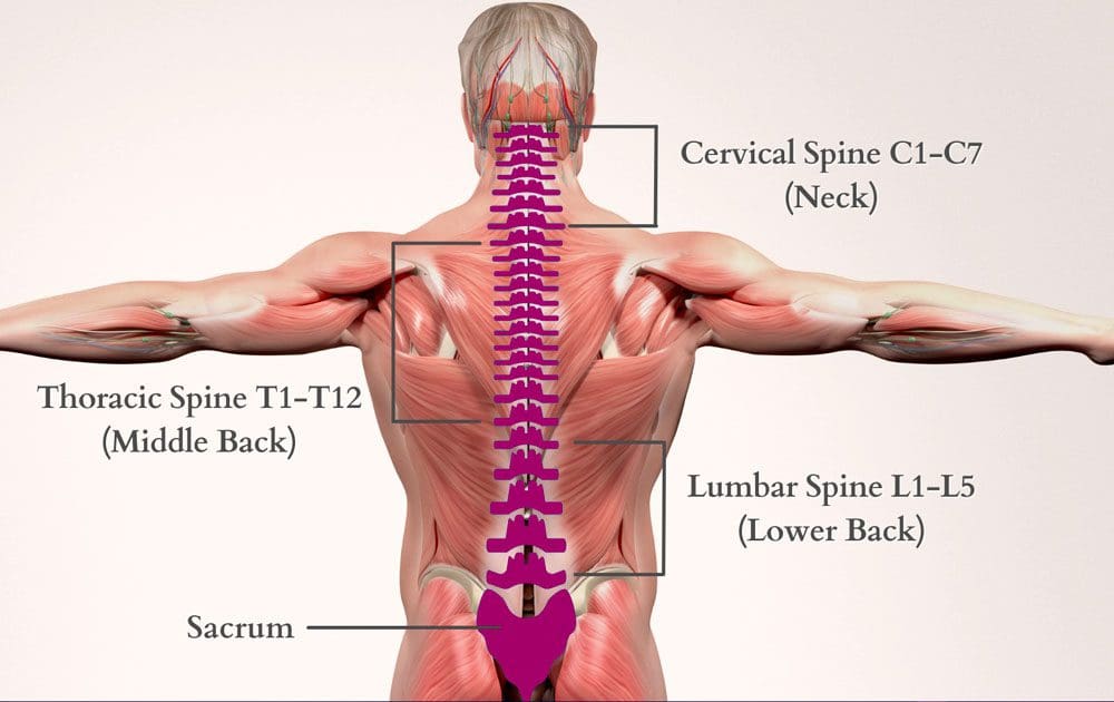
Middle back pain is usually caused by unhealthy posture, improper lifting or twisting, and minor injuries like muscle strains, sprains, and herniated discs. Thoracic herniated discs are less common than neck or low back herniations because of the thoracic vertebrae’s size and rigidity, but they do happen. Chiropractic care can treat thoracic herniated discs and prevent future episodes.
Thoracic Herniated Disc
The 12 thoracic vertebrae between the neck and the lumbar spine make up the largest and least flexible area. The rib cage adds:
Protection
Support
Stabilization of the spine
Symptoms
Herniated discs happen when the soft, gel-like layer of the shock-absorbing intervertebral disc bulges into or leaks through the disc’s tough outer layer. Due to the location, a herniated disc can cause various issues to the middle back, chest wall, and/or abdominal areas around the injured vertebrae. This displacement can cause:
Inflammation
Compression to the spinal nerves or spinal cord
Tingling
Numbness
Pain
Weakness
If the lower thoracic region is herniated, symptoms can radiate to one or both lower extremities.
Radiculopathy
If the herniation compresses a thoracic spinal nerve, it can cause radiculopathy or pain that radiates down the nerve and out from the spine into the surrounding muscles. The symptoms can present around the rib cage or upper abdominal area. A large disc herniation can compress the spinal cord inside the spinal canal. This is a condition called myelopathywhich can cause:
Numbness
Tingling
Weakness in one or both lower extremities
Sometimes bowel and bladder dysfunction
In severe cases, paralysis
Causes
Degenerative disc disease and trauma like vehicle collisions or falls are the most common causes of thoracic herniation.
Individuals between 30 and 50 are more likely to be affected.
As the body ages, the disc’s soft inner layer loses hydration, making it less effective as a shock absorber.
The tough outer layer loses elasticity, increasing the risk of disc tears.
Chiropractic Care
A chiropractor or neurologic physical therapist can personalize a herniated disc exercise treatment plan to reduce pain, improve strength and posture, and increase mobility.
Therapeutic massage can be useful in managing pain and decreasing inflammation.
Spinal epidural injections can be used with physical therapy to help manage pain and allow the body to heal independently.
Recommendations
Avoid bending, lifting, reaching, and twisting.
Apply an ice pack or cold compress for 15- to 20-minute intervals every two hours.
Sit in chairs with a firm back to support the spine.
When sleeping, place a small pillow under the head and knees to keep the spine in a neutral position to prevent pressure on the herniated region.
Avoid too much rest, which can worsen the injury.
Gentle physical activity will maintain circulation and keep the muscles strong.
Surgery
Most cases of thoracic herniation do not require surgery. Surgery could be recommended if there is intolerable pain, neurological issues, and conservative treatments are not working. A spine specialist can determine if surgery is necessary based on the injury’s size, type, and location. Spinal surgery will remove all or part of the herniated disc compressing a nerve root. Common surgical procedures include:
Court, C., E. Mansour, and C. Bouthors. “Thoracic Disc Herniation: Surgical Treatment.” Orthopaedics & Traumatology: Surgery & Research 104, no. 1 (2018). doi.org/10.1016/j.otsr.2017.04.022.
Dydyk, Alexander M, Ruben Ngnitewe Massa, and Fassil B Mesfin. “Disc Herniation – Statpearls – NCBI Bookshelf.” National Library of Medicine, January 18, 2022. www.ncbi.nlm.nih.gov/books/NBK441822/.
Yoon, Wai Weng, and Jonathan Koch. “Herniated Discs: When Is Surgery Necessary?” EFORT Open Reviews 6, no. 6 (2021): 526–30. doi.org/10.1302/2058-5241.6.210020.
IFM's Find A Practitioner tool is the largest referral network in Functional Medicine, created to help patients locate Functional Medicine practitioners anywhere in the world. IFM Certified Practitioners are listed first in the search results, given their extensive education in Functional Medicine