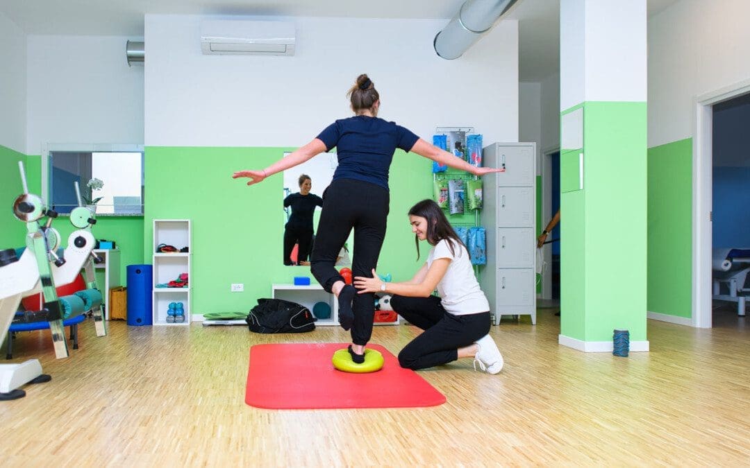
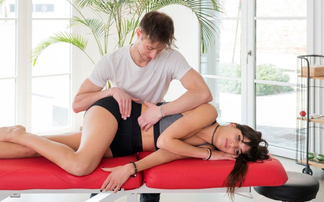
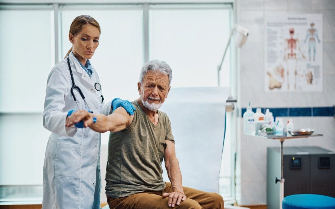
Back Clinic Injury Care Chiropractic and Physical Therapy Team. There are two approaches to injury care. They are active and passive treatment. While both can help get patients on the road toward recovery, only active treatment has a long-term impact and keeps patients moving.
We focus on treating injuries sustained in auto accidents, personal injuries, work injuries, and sports injuries and provide complete interventional pain management services and therapeutic programs. Everything from bumps and bruises to torn ligaments and back pain.
Passive Injury Care
A doctor or a physical therapist usually gives passive injury care. It includes:
Acupuncture
Applying heat/ice to sore muscles
Pain medication
It’s a good starting point to help reduce pain, but passive injury care isn’t the most effective treatment. While it helps an injured person feel better in the moment, the relief doesn’t last. A patient won’t fully recover from injury unless they actively work to return to their normal life.
Active Injury Care
Active treatment also provided by a physician or physical therapist relies on the injured person’s commitment to work. When patients take ownership of their health, the active injury care process becomes more meaningful and productive. A modified activity plan will help an injured person transition to full function and improve their overall physical and emotional wellness.
Spine, neck, and back
Headaches
Knees, shoulders, and wrists
Torn ligaments
Soft tissue injuries (muscle strains and sprains)
What does active injury care involve?
An active treatment plan keeps the body as strong and flexible as possible through a personalized work/transitional plan, which limits long-term impact and helps injured patients work toward a faster recovery. For example, in injury Medical & Chiropractic clinic’s injury care, a clinician will work with the patient to understand the cause of injury, then create a rehabilitation plan that keeps the patient active and brings them back to proper health in no time.
For answers to any questions, you may have, please call Dr. Jimenez at 915-850-0900
Progress can be challenging for individuals in post total ankle replacement surgery. How can physical therapy help in recovery and restoring leg function?
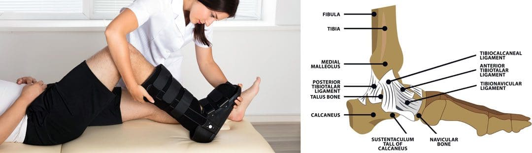
Total Ankle Replacement Post Surgery Physical Therapy
Total ankle replacement surgery is a major procedure that takes time to recover. A total ankle replacement surgery or arthroplasty can benefit individuals with chronic ankle pain or disability. This procedure can significantly improve an individual’s overall pain and function with time. Physical therapy is essential to regaining movement in the ankle and restoring full mobility. A physical therapist will work with the individual to control pain and swelling, restore the ankle’s range of motion, train on walking gait and balance, and rebuild strength in the leg. This will help maximize the chances of a successful outcome after surgery.
Total Ankle Replacement
The ankle joint is the section of the lower leg where the shinbone/tibia meets the talus bone on the top of the foot. What can happen is the slippery surface/articular cartilage that coats the ends of these bones begins to thin or deteriorate. As the deterioration progresses, it can lead to significant pain, disability, and difficulty walking. (Cleveland Clinic. 2021) This is where a specialist may recommend total ankle replacement for the best results. Various conditions can be helped by this procedure, including:
During an ankle replacement procedure, an orthopedic surgeon removes the damaged ends of the tibia and talus bones and replaces them with an artificial covering. A polyethylene component is also secured between the two structures to support the smooth movement of the new joint endings. (Massachusetts General Hospital. N.D.) Following the procedure, individuals are typically placed in a protective boot or splint. The healthcare provider will recommend staying off the leg for 4 to 8 weeks to allow healing.
Physical Therapy
Outpatient physical therapy is usually initiated several weeks after the ankle operation. (UW Health Orthopedics and Rehabilitation. 2018) Physical therapy can last for five months or more, depending on the severity of the condition and injury. The physical therapist will focus on different areas to get the best results. (Cort D. Lawton et al., 2017)
Pain and Swelling Control
Post-operative pain and swelling are normal after a total ankle replacement. It is not unusual for an ankle to be swollen for even six to 12 months after the operation. (UW Health Orthopedics and Rehabilitation. 2018) The surgeon will normally prescribe medication to help manage discomfort early on, and physical therapy also plays an important role in addressing the symptoms. Treatments used can include:
Electrical stimulation – mild electrical pulses applied to the muscles.
Ice
Vasopneumatic compression, where an inflatable sleeve is used to create pressure around the area, is commonly utilized at the beginning of physical therapy to reduce pain or swelling.
Other modalities, such as stretching and targeted exercises, are combined with other treatments.
Range of Motion
Early after the procedure, the ankle will be very stiff and tight. This is due to several factors, including the inflammation and swelling after surgery and the time spent immobilized in a boot.
The physical therapist will employ various techniques to improve the ankle joint’s range of motion to rotate and flex.
The physical therapist may employ passive stretching induced by an outside force such as the therapist or a resistance band) to help improve mobility.
After multiple weeks of reduced movement and lack of bearing any weight on the ankle, the muscles that surround the ankle have often atrophied/weakened, which can impact balance.
When the individual can begin placing weight on the leg, the therapist will apply proprioceptive/sense of body position training to improve overall stability. (UW Health Orthopedics and Rehabilitation. 2018)
Balance exercises will be added to the home program and will progress from week to week.
Strength
The muscles in the leg, ankle, and foot become weak from the surgery and the time spent in a splint or boot. These structures have a significant role in balance, the ability to stand, walk, and go up or down the stairs.
Regaining the strength and power of these muscles is a critical goal of rehabilitation.
In the first weeks, the physical therapist will focus on gentle strengthening exercises.
Isometrics lightly activate the muscles but avoid irritating the surgical site.
As time passes and weight-bearing is allowed, these gentle moves are replaced with more challenging ones, like resistance bands and standing exercises, to accelerate strength gains.
Lawton, C. D., Butler, B. A., Dekker, R. G., 2nd, Prescott, A., & Kadakia, A. R. (2017). Total ankle arthroplasty versus ankle arthrodesis-a comparison of outcomes over the last decade. Journal of orthopaedic surgery and research, 12(1), 76. doi.org/10.1186/s13018-017-0576-1
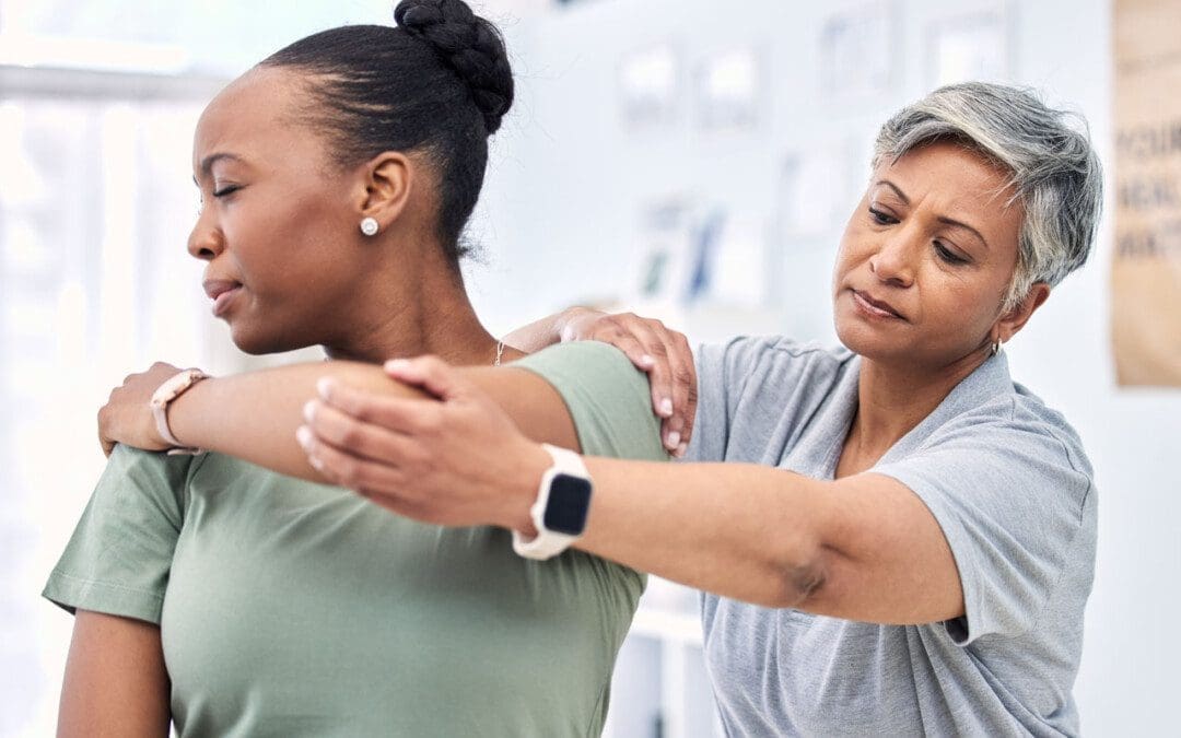
For individuals having difficulty moving or functioning normally due to injury, surgery, or illness, can a chiropractic and physical therapy team help expedite recovery?
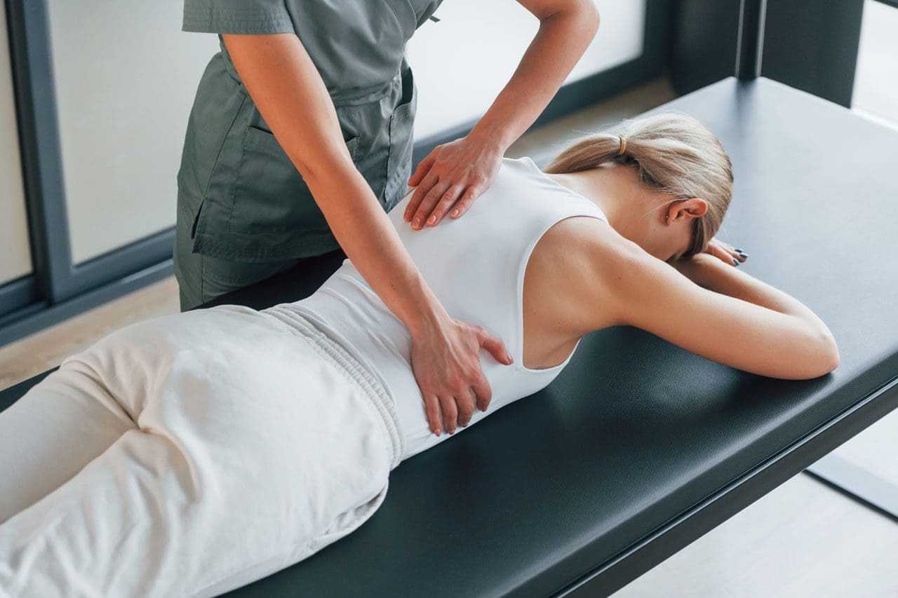
Friction Massage
Individuals may develop scar tissue or tissue adhesions that limit normal motion after injury or surgery. A pain management team may use various treatments and modalities and may incorporate friction massage as part of a rehabilitation treatment plan. Friction massage, also known as transverse friction or cross friction massage, is a technique used to help improve scar tissue and adhesion mobility to move better and decrease the negative effects. The therapist uses their fingers to massage the scar in a direction that is at right angles to the scar line. It is a specialized technique that breaks up tissue adhesions that are limiting normal movement in the skin and underlying tissues. (Haris Begovic, et al., 2016)
Scar Tissue and Adhesions
For individuals who require surgery due to an injury or an orthopedic condition, their doctor will cut into the skin, tendons, and muscle tissue during the operation. Once sutured and healing has begun, scar tissue forms. Healthy tissue is made up of collagen that is comprised of cells that are arranged in a regular pattern. Healthy collagen is strong and can resist forces when tissues are pulled and stretched. (Paula Chaves, et al., 2017)
During the healing process after an injury, the collagen cells are laid down in a haphazard pattern and form scar tissue. The random accumulation of cells becomes tight and does not react well to tension and stretching forces. (Qing Chun, et al., 2016) The body can form scar tissue after a soft tissue injury, like a muscle or tendon strain. (Qing Chun, et al., 2016)
If a muscle or tendon gets strained the body will generate new collagen during the healing. The new collagen is laid down in a random fashion, and scar tissue or tissue adhesions can form that can limit the normal range of motion. Healthy tissue stretches and glides as the body moves. Scar tissue is rigid. At the site of the scar tissue, there can be some movement, but it is tight, less pliable, and can be painful. If scar tissue or adhesions are limiting motion, cross-friction massage can improve tissue gliding and sliding. This process is referred to as remodeling.
Massage Objectives
The objectives and goals of friction massage to adhesions or scar tissue may include:
Stimulation of nerve fibers to decrease and relieve pain.
The entire area of scar tissue or adhesion should be treated.
If the scar tissue is in a muscle, it should be relaxed.
If the scar tissue is in a tendon sheath, that tendon should be slightly stretched during the procedure.
The therapist places two or three fingers over the scar or adhesion and moves their fingers perpendicular to the scar to smooth the collagen fibers down.
The fingers and underlying tissues move together.
The massage should feel deep and uncomfortable but not painful.
There may be some pain, but should remain within the individual’s tolerance.
If the massage is too painful, less pressure may be used.
After several minutes the therapist will assess the tissue mobility.
Specific stretches may be done to elongate the scar tissue or adhesions.
At-home exercises and stretches may be prescribed to maintain flexibility.
Contraindications
There are situations where friction massage should not be used and can include: (Paula Chaves, et al., 2017)
Around an active open wound.
If there is a bacterial infection.
Areas with decreased sensation.
If calcification is present in the muscle or tendon tissue.
The therapist will explain the procedure and inform of the goals and risks associated with it.
Adhesive capsulitis in the shoulder/frozen shoulder.
Joint contracture.
Ligament tears.
Scar tissue buildup after surgery or trauma.
Friction massage is a popular technique used in physical therapy, but some research suggests it is not any more effective than other rehabilitation techniques. One study found that static stretches and exercises were more effective than massage in improving tissue length and strength in uninjured soccer players. Other studies have supported this, but individuals may find that the massage helps improve injured tissues’ movement as well. (Mohammed Ali Fakhro, et al. 2020)
The main goal of any treatment in physical therapy is to help the individual regain movement and flexibility. Friction massage, combined with targeted stretches and exercises, can help individuals expedite recovery and get back to normal.
Chiropractic Care After Accidents and Injuries
References
Begovic, H., Zhou, G. Q., Schuster, S., & Zheng, Y. P. (2016). The neuromotor effects of transverse friction massage. Manual therapy, 26, 70–76. doi.org/10.1016/j.math.2016.07.007
Chaves, P., Simões, D., Paço, M., Pinho, F., Duarte, J. A., & Ribeiro, F. (2017). Cyriax’s deep friction massage application parameters: Evidence from a cross-sectional study with physiotherapists. Musculoskeletal science & practice, 32, 92–97. doi.org/10.1016/j.msksp.2017.09.005
Chun, Q., ZhiYong, W., Fei, S., & XiQiao, W. (2016). Dynamic biological changes in fibroblasts during hypertrophic scar formation and regression. International wound journal, 13(2), 257–262. doi.org/10.1111/iwj.12283
Fakhro, M. A., Chahine, H., Srour, H., & Hijazi, K. (2020). Effect of deep transverse friction massage vs stretching on football players’ performance. World journal of orthopedics, 11(1), 47–56. doi.org/10.5312/wjo.v11.i1.47
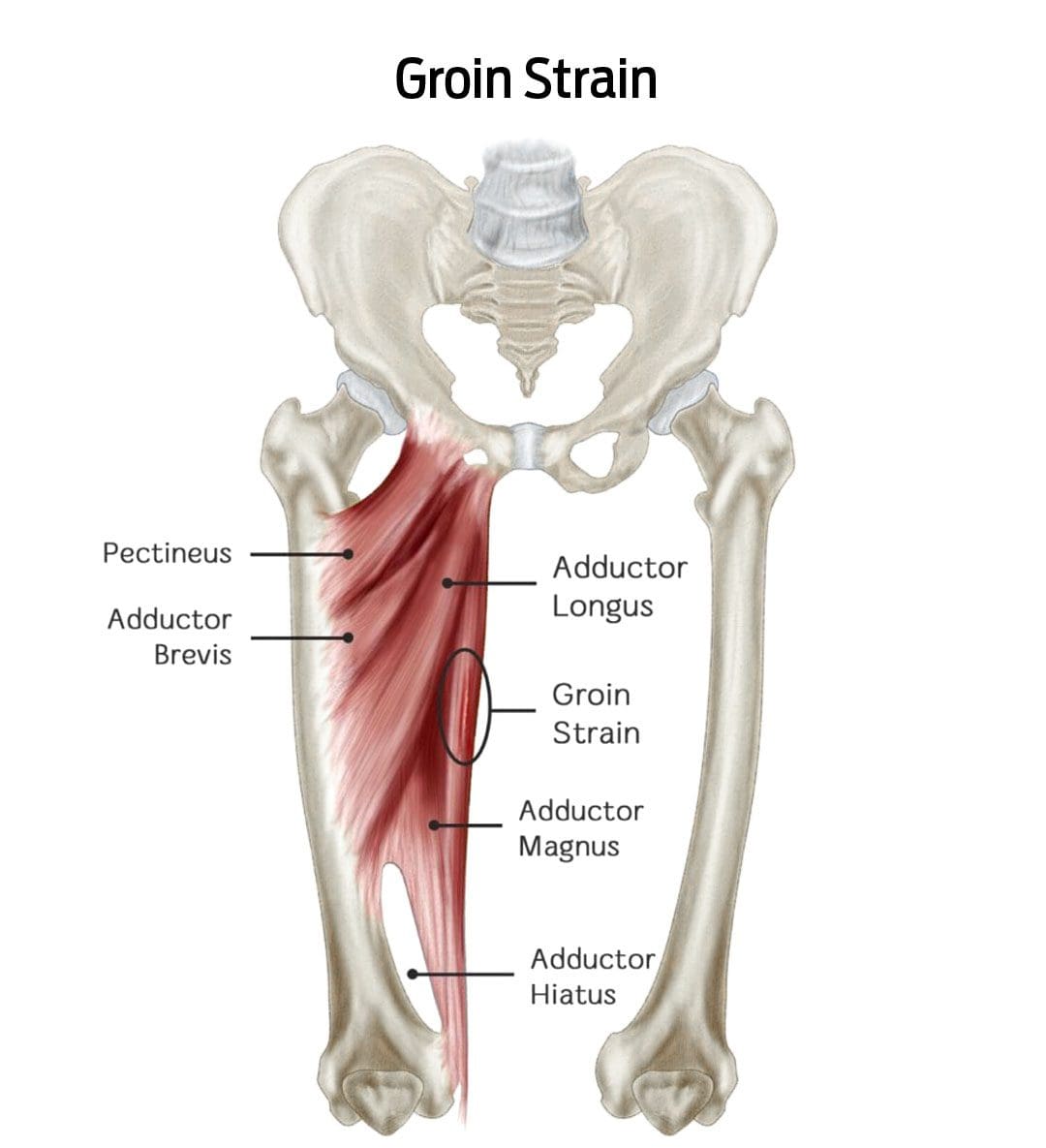
When a groin strain injury happens, can knowing the symptoms help in the diagnosis, treatment, and recovery times?
Groin Strain Injury
A groin strain is an injury to an inner thigh muscle. A groin pull is a type of muscle strain affecting the adductor muscle group (the muscles help pull the legs apart). (Parisa Sedaghati, et al., 2013) The injury is caused when the muscle is stretched beyond its normal range of motion, creating superficial tears. Severe strains can tear the muscle in two. (Parisa Sedaghati, et al., 2013)
A groin muscle pull causes pain and tenderness that worsens when squeezing the legs together.
There may also be swelling or bruising in the groin or inner thigh.
An uncomplicated groin pull takes four to six weeks to heal with proper treatment. (Andreas Serner, et al., 2020)
Symptoms
A groin pull can be painful, interfering with walking, navigating stairs, and/or driving a car. In addition to pain, other symptoms around the injured area include: (Parisa Sedaghati et al., 2013)
A popping sound or snapping sensation when the injury occurs.
Increased pain when pulling the legs together.
Redness
Swelling
Bruising of the groin or inner thigh.
Groin pulls are graded by severity and how much they impact mobility:
Grade 1
Mild discomfort but not enough to limit activities.
Grade 2
Moderate discomfort with swelling or bruising that limits running and/or jumping.
Grade 3
Severe injury with significant swelling and bruising can cause pain while walking and muscle spasms.
Signs of a severe groin strain
Difficulty walking
Groin pain while sitting or resting
Groin pain at night
A healthcare provider should see severe groin pulls because the muscle may have ruptured or be on the verge of rupturing.
In severe cases, surgery is necessary to reattach the torn ends.
Groin pulls are sometimes accompanied by a stress fracture of the pubis/forward-facing pelvic bones, which can significantly extend healing and recovery time. (Parisa Sedaghati et al., 2013)
Causes
Groin pulls are often experienced by athletes and individuals who play sports where they must stop and change directions quickly, placing excessive strain on the adductor muscles. (Parisa Sedaghati et al., 2013) The risk is increased in individuals who: (T. Sean Lynch et al., 2017)
Have weak hip abductor muscles.
Are not in adequate physical condition.
Have a previous groin or hip injury.
Pulls can also occur from falls or extreme activities without the proper conditioning.
Diagnosis
A healthcare provider will perform a thorough investigation to confirm the diagnosis and characterize the severity. This involves: (Juan C. Suarez et al., 2013)
Medical History Review
This includes any previous injuries and specifics about where and when the symptoms started.
Physical Examination
This involves palpating – lightly touching and pressing the groin region and manipulating the leg to understand better where and how extensive the injury is.
Imaging Studies
Ultrasound or X-rays.
If a muscle rupture or fracture is suspected, an MRI scan may be ordered to visualize soft tissue injuries and stress fractures better.
Differential Diagnosis
Certain conditions can mimic a groin pull and require different treatments. These include: (Juan C. Suarez, et al., 2013)
Sports Hernia
This type of inguinal hernia occurs with sports and work injuries.
It causes a portion of the intestine to pop through a weakened muscle in the groin.
Hip Labral Tear
This is a tear in the cartilage ring of the labrum outside the rim of the hip joint socket.
Hip Osteoarthritis
This is the wear-and-tear form of arthritis that can present with groin pain symptoms.
Osteitis Pubis
This is inflammation of the pubic joint and surrounding structures, usually caused by the overuse of the hip and leg muscles.
Referred Groin Pain
This nerve pain originates in the lower back, often due to a pinched nerve, but is felt in the groin.
Treatment
Beginning treatment is conservative and includes rest, ice application, physical therapy, and prescribed gentle stretching and exercises.
Individuals may need crutches or a walking device to reduce pain and prevent further injury if the pain is significant. (Andreas Serner, et al., 2020)
Physical therapy will be a part of the treatment plan.
Over-the-counter pain medications like Tylenol/acetaminophen or Advil/ibuprofen can help with pain relief short term.
If there is severe pain from a grade 3 injury, prescription medications may be used for a short period to help minimize pain. (Andreas Serner, et al., 2020)
Sedaghati, P., Alizadeh, M. H., Shirzad, E., & Ardjmand, A. (2013). Review of sport-induced groin injuries. Trauma monthly, 18(3), 107–112. doi.org/10.5812/traumamon.12666
Serner, A., Weir, A., Tol, J. L., Thorborg, K., Lanzinger, S., Otten, R., & Hölmich, P. (2020). Return to Sport After Criteria-Based Rehabilitation of Acute Adductor Injuries in Male Athletes: A Prospective Cohort Study. Orthopaedic journal of sports medicine, 8(1), 2325967119897247. doi.org/10.1177/2325967119897247
Lynch, T. S., Bedi, A., & Larson, C. M. (2017). Athletic Hip Injuries. The Journal of the American Academy of Orthopaedic Surgeons, 25(4), 269–279. doi.org/10.5435/JAAOS-D-16-00171
Suarez, J. C., Ely, E. E., Mutnal, A. B., Figueroa, N. M., Klika, A. K., Patel, P. D., & Barsoum, W. K. (2013). Comprehensive approach to the evaluation of groin pain. The Journal of the American Academy of Orthopaedic Surgeons, 21(9), 558–570. doi.org/10.5435/JAAOS-21-09-558
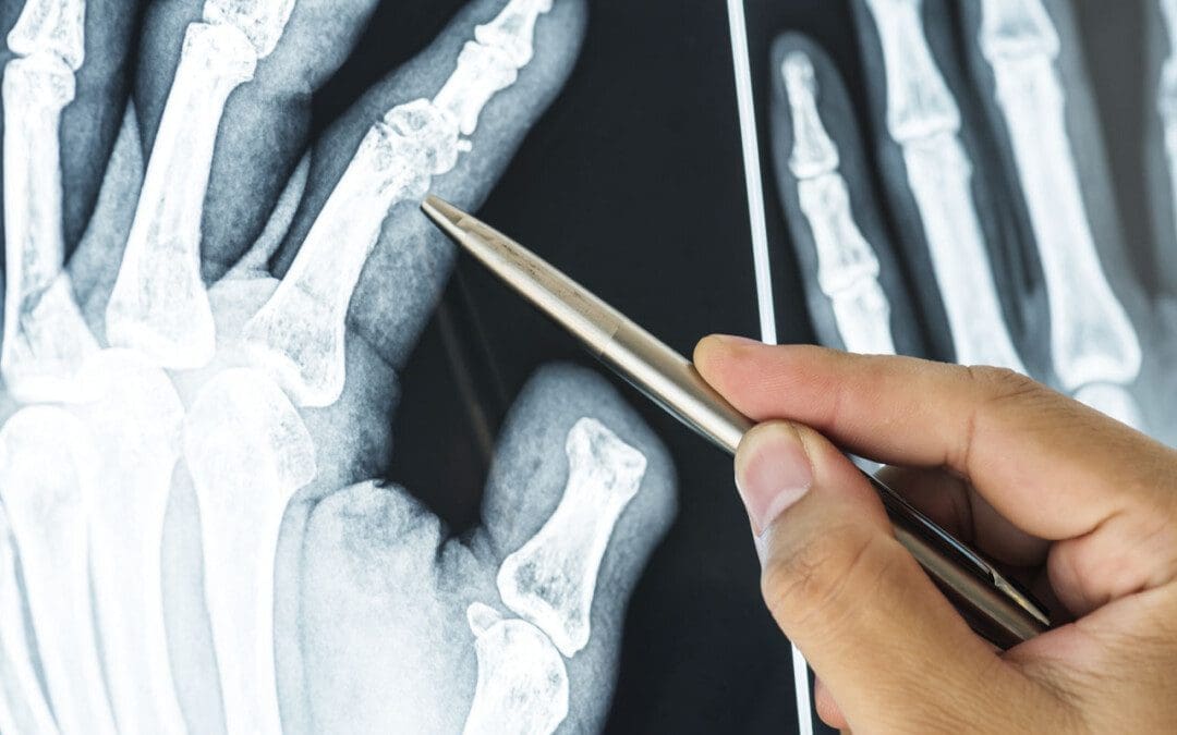
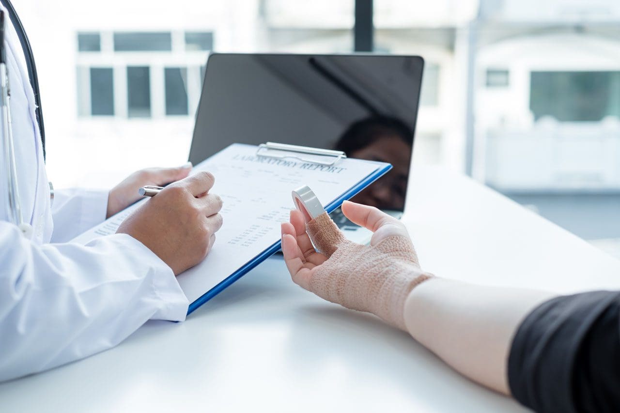
Finger sprains and dislocations are common hand injuries that can happen during work, physical/sports activities, or in automobile collisions and accidents. Can recognizing the symptoms help in developing an effective treatment strategy?
Finger Sprains and Dislocations
Finger sprains and dislocations are common injuries of the hand that cause pain and swelling.
A sprain happens when the finger tissue that supports a joint gets stretched beyond its limits in a way that stresses the ligaments and tendons.
The ligament tissue can be partially or completely torn. If the damage is bad enough, the joint comes apart.
This is a dislocation – A dislocation happens when the joint in the finger gets shifted out of its normal position.
Both injuries can cause pain and stiffness in the finger and hand.
Sprains
Finger sprains can happen any time the finger bends in an awkward or unusual way. This can happen from falling on the hand or getting hurt when engaged in physical activities like sports or household chores. Sprains can occur in any of the knuckle joints in the finger. However, most commonly, the joint in the middle of the finger gets sprained. It’s known as the proximal interphalangeal or PIP joint. (John Elfar, Tobias Mann. 2013) Symptoms of a finger sprain can include:
Other treatments to help a sprained finger include:
Elevate the hand if swelling and inflamed.
Gentle finger exercises/movements to prevent stiffness.
Icing the injured finger.
Take an anti-inflammatory medication.
Individuals who have not broken bones or dislocated the joint will probably be able to move their finger in about a week. A doctor will set a timeline for when to start using the finger normally.
Individuals who sprain their finger that feels swollen and stiff for longer than a few weeks are recommended to consult a doctor or specialist.
Thumb sprains and finger sprains in children may need to be splinted or taped for longer periods, as the ligament is not fully developed or as strong, which could lead to a tear.
Dislocations
A finger dislocation is a more severe injury involving the ligament, joint capsule, cartilage, and other tissues that causes misalignment of the finger. The ligaments and the joint capsule get torn when a joint is dislocated. The joint needs to be reset, which can be a simple process, or in severe cases, patients may need to be placed under anesthesia or undergo surgery to reset the joint properly.
In these cases, tendons or other tissues might be preventing the joint from getting into position.
Putting the finger back into the right position is known as”reduction.” Once reduced, the finger needs to be splinted.
Individuals also need an X-ray to ensure the joint is lined up correctly and that any bones were not broken or fractured when they sustained the injury. (James R. Borchers, Thomas M. Best. 2012)
Once reset, caring for a dislocated finger is basically the same as a sprained finger. Using ice on the finger, keeping the hand elevated to reduce swelling.
Elfar, J., & Mann, T. (2013). Fracture-dislocations of the proximal interphalangeal joint. The Journal of the American Academy of Orthopaedic Surgeons, 21(2), 88–98. doi.org/10.5435/JAAOS-21-02-88
OrthoInfo from the American Academy of Orthopaedic Surgeons. (2022) Hand fractures.
Hung, C. Y., Varacallo, M., & Chang, K. V. (2023). Gamekeeper’s Thumb. In StatPearls. StatPearls Publishing.
OrthoInfo from the American Academy of Orthopaedic Surgeons. (2022) Finger fractures.
Borchers, J. R., & Best, T. M. (2012). Common finger fractures and dislocations. American family physician, 85(8), 805–810.
As the body grows older the ability to live life to the fullest can be difficult. Can using natural biologics help enhance the body’s natural ability to heal?
Natural Biologics
Though sometimes a necessary treatment option, surgical procedures can be the first line of treatment introduced to patients. Natural biologics is a less invasive alternative that can eliminate hospitalizations and expedite recovery. (Riham Mohamed Aly, 2020)
What Are They?
The body is born with components to initiate healing and recovery. These components include:
Cells
Cytokines
Proteins
Collagens
Elastin
Hyaluronic acid
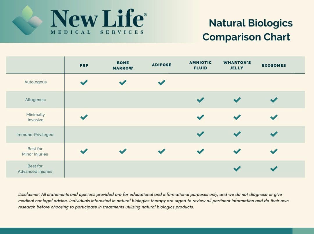
At the time of birth, these components are in abundance but decrease as the body ages. This is why children recover from injuries quicker than adults. Recovery for adults can be slower from a decrease in these natural healing components. The objective of natural biologics treatments is to increase the healing components by reintroducing the body’s own components – autologous – or by bringing in new components – allogeneic – from a donor. (National Institutes of Health 2016) Choosing between the two options depends on an individual’s age and health, as those who are older or in poor physical health may experience complications from inferior component amounts.
Healing components derived from donor sources can show more promise, as treatments are usually acquired from discarded birth tissues at delivery.
Birth tissues are rich in healing components, containing the most abundant collection of natural healing elements.
It’s important to note that there is no harm to the mother or the baby from the obtained tissue products.
Platelet-rich plasma is cultivated by drawing an individual’s blood and spinning it in a centrifuge to separate the plasma.
The resulting liquid is reinjected into the injured area to generate a healing environment.
This form of natural biologics is effective for individuals with minor injuries that can be repaired easily.
This process is not as effective for older individuals who already have a reduction in natural healing components.
Lifestyle factors such as smoking, unhealthy diet, and alcohol/substance abuse can decrease the effectiveness of PRP treatments.
Bone Marrow Aspirate
This is an invasive, painful process that begins by putting a patient under anesthesia and drilling into the bone to extract the marrow. (American Cancer Society, 2023)
Like PRP, success depends on the individual’s age, health, and lifestyle.
Invasive procedures like this have a higher probability of infection and require a long-term recovery period.
Adipose-Derived Stem Cells
Adipose tissue/fat treatments are collected through a procedure that resembles the process of liposuction.
The procedure is done under general anesthesia and is an invasive process.
The treatment’s success depends on the individual’s health, age, and lifestyle.
There is more risk of infection when choosing this procedure and a long-term recovery period.
Allogeneic Treatment
Donor-based regenerative cells.
Amniotic Fluid Therapy
Amniotic fluid contains various growth factors, cytokines, and anti-inflammatory proteins that may promote tissue repair, reduce inflammation, and stimulate cellular regeneration. (Petra Klemmt. 2012)
Collected at the time of birth, this therapy is an ideal treatment for individuals who have sustained injuries that affect day-to-day functionality.
Physicians and clinicians are utilizing amniotic fluid therapy to treat many conditions, from orthopedic to wound care.
Amniotic fluid is collected at the time of birth and is abundant with increased healing components compared to autologous sources.
Amniotic fluid is immune-privileged (limits or suppresses immune response) and the risk of rejection is rare.
These therapies are usually done in a physician’s office with minimal downtime after treatment.
Wharton’s Jelly
Wharton’s jelly is derived from the umbilical cord at the time of birth and is primarily composed of a gel substance made up of hyaluronic acid and a network of collagen fibers.
Believed to contain a population of mesenchymal stem cells that have the capacity to differentiate into various cell types, and other secreted growth factors and cytokines. (F. Gao, et al., 2016)
It is considered the most valuable source to enhance the healing of various tissues, including bone, cartilage, skin, and nerve tissue.
It is immune-privileged with little risk of rejection and minimal if any, recovery time after an in-office treatment.
Exosomes
Exosomes are small, membrane-bound vesicles that play a role in intercellular communication within the body. (Carl Randall Harrell, et al., 2019)
They contain a variety of bioactive molecules, including proteins, lipids, nucleic acids (like RNA), and signaling molecules.
They serve as vehicles for transferring the signaling molecules from one cell to another, allowing cells to influence the behavior and function of neighboring or distant cells.
They can be collected or isolated from various biological fluids and cell cultures through specialized techniques but are most robust when collected at birth.
The exosomes within the umbilical cord are utilized for tissue repair and regeneration, signaling the cells to promote:
Proliferation – increase in the number of cells through cell division.
Differentiation – the transformation of unspecialized cells into specialized cells.
Tissue healing in damaged or injured areas.
Exosomes from the umbilical cord are immune-privileged with minimal risk of rejection.
Treatments are ideal for increasing cell communication and initiating repair when paired with another source of allogeneic therapy like amniotic fluid or Wharton’s Jelly.
Choosing which natural biologics therapy is the best is different for everyone. When selecting a treatment, it is essential for individuals to consult their primary healthcare provider to determine which application will have optimal results.
Is Motion Key To Healing?
References
Aly R. M. (2020). Current state of stem cell-based therapies: an overview. Stem cell investigation, 7, 8. doi.org/10.21037/sci-2020-001
Mazini, L., Rochette, L., Admou, B., Amal, S., & Malka, G. (2020). Hopes and Limits of Adipose-Derived Stem Cells (ADSCs) and Mesenchymal Stem Cells (MSCs) in Wound Healing. International journal of molecular sciences, 21(4), 1306. doi.org/10.3390/ijms21041306
Klemmt P. (2012). Application of amniotic fluid stem cells in basic science and tissue regeneration. Organogenesis, 8(3), 76. doi.org/10.4161/org.23023
Sabapathy, V., Sundaram, B., V M, S., Mankuzhy, P., & Kumar, S. (2014). Human Wharton’s Jelly Mesenchymal Stem Cells plasticity augments scar-free skin wound healing with hair growth. PloS one, 9(4), e93726. doi.org/10.1371/journal.pone.0093726
Gao, F., Chiu, S. M., Motan, D. A., Zhang, Z., Chen, L., Ji, H. L., Tse, H. F., Fu, Q. L., & Lian, Q. (2016). Mesenchymal stem cells and immunomodulation: current status and future prospects. Cell death & disease, 7(1), e2062. doi.org/10.1038/cddis.2015.327
Harrell, C. R., Jovicic, N., Djonov, V., Arsenijevic, N., & Volarevic, V. (2019). Mesenchymal Stem Cell-Derived Exosomes and Other Extracellular Vesicles as New Remedies in the Therapy of Inflammatory Diseases. Cells, 8(12), 1605. doi.org/10.3390/cells8121605
When individuals experience a neuromusculoskeletal injury strain, can following basic pulled muscle treatment protocols help in healing and a full recovery?
Pulled Muscle Treatment
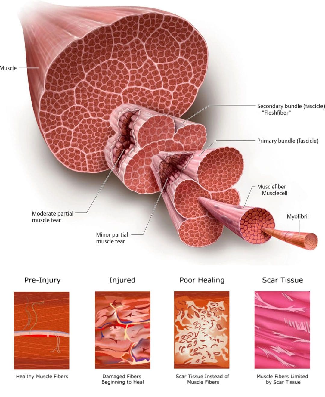
A pulled muscle or muscle strain occurs when a muscle is stretched beyond its ability resulting in discomfort symptoms and mobility issues. Microscopic tears can occur within the muscle fibers potentially worsening the injury. This type of injury usually causes mild to severe pain, bruising, and immobility, and nerve injuries can develop as well. Common muscle strains include:
Pulled hamstrings
Groin strains
Pulled abdominal muscles
Calf strains
Pulled muscle treatment requires patience to promote proper healing and restoration of optimal function.
Individuals need to focus on the different stages of healing.
Gradually increase activity levels as the body allows to prevent stiffness and atrophy which can cause complications.
Symptoms
The usual symptoms of this type of injury include:
Pain
Limited mobility
Muscle spasms
Swelling
Bruising
Often individuals will feel a sudden grabbing or tearing sensation and are then unable to continue the activity.
Can limit the ability to perform certain activities.
May have moderate swelling and bruising.
Grade III
Severe injury that can cause significant pain.
Muscle spasms.
Swelling.
Significant bruising.
Basic Treatment Protocols
Most pulled muscle strain injuries heal with simple treatment. Following the right steps can ensure an expedited recovery. In the early stages after the injury, there is a balance between doing too much or not enough. The amount of activity an individual will be able to do, and the time required for recovery depends on the severity of the injury. Here are some guidelines in the right direction.
Rest
Rest is recommended for the early recovery stage.
Depending on the severity of the injury this could last from one to five days.
Immobilization is usually not necessary, and not moving at all can lead to muscle and joint stiffness.
To avoid injuries make sure the muscles are not over-exerted.
Gradually increase activity levels when starting an exercise program to build endurance.
Properly Warming Up
Warming up before taking on physical activities will help loosen the muscles and prevent injuries.
Beginning work or exercise with stiff muscles can lead to an increased chance of strain.
Studies have shown that temperature can influence the stiffness of a muscle. (K. W. Ranatunga. 2018)
Maintaining body and muscle warmth helps prevent injury and re-injury.
Injuries and Chiropractic: The Road To Recovery
References
Hospital for Special Surgery, Muscle Strain: What You Need to Know About Pulled Muscles.
Kary J. M. (2010). Diagnosis and management of quadriceps strains and contusions. Current reviews in musculoskeletal medicine, 3(1-4), 26–31. doi.org/10.1007/s12178-010-9064-5
Malanga, G. A., Yan, N., & Stark, J. (2015). Mechanisms and efficacy of heat and cold therapies for musculoskeletal injury. Postgraduate medicine, 127(1), 57–65. doi.org/10.1080/00325481.2015.992719
Mair, S. D., Seaber, A. V., Glisson, R. R., & Garrett, W. E., Jr (1996). The role of fatigue in susceptibility to acute muscle strain injury. The American journal of sports medicine, 24(2), 137–143. doi.org/10.1177/036354659602400203
Ranatunga K. W. (2018). Temperature Effects on Force and Actin⁻Myosin Interaction in Muscle: A Look Back on Some Experimental Findings. International journal of molecular sciences, 19(5), 1538. doi.org/10.3390/ijms19051538
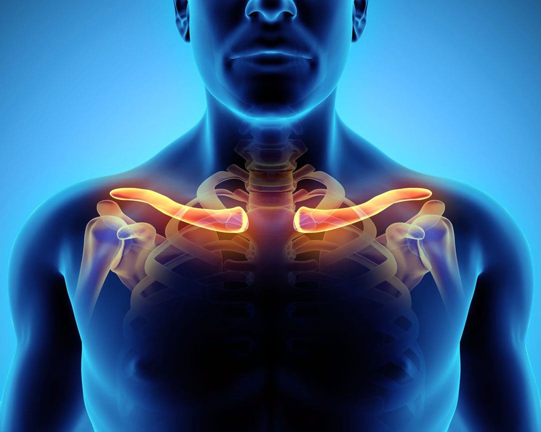
For individuals with a broken collarbone, can conservative treatment help in the rehabilitation process?
Broken Collarbone
Broken collarbones are very common orthopedic injuries that can occur in any age group. Also known as the clavicle, it is the bone over the top of the chest, between the breastbone/sternum and the shoulder blade/scapula. The clavicle can be easily seen because only skin covers a large part of the bone. Clavicle fractures are extremely common, and account for 2% – 5% of all fractures. (Radiopaedia. 2023) Broken collarbones occur in:
Babies – usually during birth.
Children and adolescents – because the clavicle does not fully develop until the late teens.
Athletes – because of the risks of being hit or falling.
Through various types of accidents and falls.
The majority of broken collarbones can be treated with nonsurgical treatments, usually, with a sling to let the bone heal and physical therapy and rehabilitation.
Sometimes, when clavicle fractures are significantly shifted out of alignment, surgical treatment may be recommended.
There are treatment options that should be discussed with an orthopedic surgeon, physical therapist, and/or a chiropractor.
A broken collarbone is not more serious than other broken bones.
Once the broken bone heals, most individuals have a full range of motion and can return to the activities before the fracture. (Johns Hopkins Medicine. 2023)
Types
Broken clavicle injuries are separated into three types depending on the location of the fracture. (Radiopaedia. 2023)
Mid-Shaft Clavicle Fractures
These occur in the central area which can be a simple crack, separation, and/or fractured into many pieces.
Multiple breaks – segmental fractures.
Significant displacement – separation.
Shortened length of the bone.
Distal Clavicle Fractures
These happen close to the end of the collarbone at the shoulder joint.
This part of the shoulder is called the acromioclavicular/AC joint.
Distal clavicle fractures can have similar treatment options as an AC joint injury.
Medial Clavicle Fractures
These are less common and often related to injury to the sternoclavicular joint.
The sternoclavicular joint supports the shoulder and is the only joint that connects the arm to the body.
Growth plate fractures of the clavicle can be seen into the late teens and early 20s.
The bruising can extend down to the chest and armpit.
Numbness and tingling down the arm.
Deformity of the collarbone.
In addition to swelling, some individuals may have a bump in the place where the fracture occurred.
It can take several months for this bump to fully heal, but this is normal.
If the bump appears inflamed or irritated, inform a healthcare provider.
Clavicular Swelling
When the sternoclavicular joint swells up or gets bigger, it is referred to as clavicular swelling.
It is commonly caused by trauma, disease, or an infection that affects the fluid found in the joints. (John Edwin, et al., 2018)
Diagnosis
At the healthcare clinic or emergency room, an X-ray will be obtained to assess for the specific type of fracture.
They will perform an examination to ensure the nerves and blood vessels surrounding the broken collarbone are unsevered.
The nerves and vessels are rarely injured, but in severe cases, these injuries can occur.
Treatment
Treatment is accomplished either by allowing the bone to heal or by surgical procedures to restore the proper alignment. Some common treatments for broken bones are not used for clavicle fractures.
For example, casting a broken collarbone is not done.
In addition, resetting the bone or a closed reduction is not done because there is no way to hold the broken bone in proper alignment without surgery.
If surgery is an option the healthcare provider looks at the following factors: (UpToDate. 2023)
Location of Fracture and Degree of Displacement
Nondisplaced or minimally displaced fractures are usually managed without surgery.
Age
Younger individuals have an increased ability to recover from fractures without surgery.
Shortening of the Fracture Fragment
Displaced fractures can heal, but when there is a pronounced shortening of the collarbone, surgery is probably necessary.
Other Injuries
Individuals with head injuries or multiple fractures can be treated without surgery.
Patient Expectations
When the injury involves an athlete, heavy job occupation, or the arm is the dominant extremity, there can be more reason for surgery.
Dominant Arm
When fractures occur in the dominant arm, the effects are more likely to be noticeable.
The majority of these fractures can be managed without surgery, but there are situations where surgery can produce better results.
Supports for Non-surgical Treatment
A sling or figure-8 clavicle brace.
The figure-8 brace has not been shown to affect fracture alignment, and many individuals generally find a sling more comfortable. (UpToDate. 2023)
Broken collarbones should heal within 6–12 weeks in adults
3–6 weeks in children
Younger patients are usually back to full activities before 12 weeks.
The pain usually subsides within a few weeks. (UpToDate. 2023)
Immobilization is rarely needed beyond a few weeks, and with a doctor’s clearance light activity and gentle motion rehabilitation usually begins.
Edwin, J., Ahmed, S., Verma, S., Tytherleigh-Strong, G., Karuppaiah, K., & Sinha, J. (2018). Swellings of the sternoclavicular joint: review of traumatic and non-traumatic pathologies. EFORT open reviews, 3(8), 471–484. doi.org/10.1302/2058-5241.3.170078
IFM's Find A Practitioner tool is the largest referral network in Functional Medicine, created to help patients locate Functional Medicine practitioners anywhere in the world. IFM Certified Practitioners are listed first in the search results, given their extensive education in Functional Medicine