L-glutamate is one of the main excitatory neurotransmitters in the human brain and it plays an essential role in practically all activities of the nervous system. In the following article, we will discuss the general principles of L-glutamate signaling in the brain. Then, we will demonstrate this scheme by describing the different pools of extracellular glutamate, including the synaptic, the perisynaptic, and the extrasynaptic, resulting from vesicular and non-vesicular sources or abnormally located glutamate receptors outside of synapses as well as discuss their possible physiological functions in the human brain. �
Contents
Glutamate Signaling in the Brain
According to research studies, the human brain has about a 6 to 7 ?mol/g wet weight of L-glutamate. L-glutamate, together with glutamine, is one of the most abundant free amino acids in the central nervous system (CNS). More than five decades ago, several research studies demonstrated that L-glutamate has an excitatory response on nerve cells. Since then, its role as an excitatory neurotransmitter as well as its cerebral metabolism has been evaluated in numerous research studies. �
L-glutamate is commonly found throughout synaptic vesicles in the presynaptic terminal through the process of vesicular glutamate transporters. Additionally, several of the L-glutamate in the vesicles may develop by a vesicle-associated aspartate amino-transferase from 2-oxoglutarate utilizing L-aspartate as the amino group donor. During the depolarization of the presynaptic membrane, L-glutamate is released into the synaptic cleft and connects to ionotropic glutamate receptors, known as iGluRs, at the postsynaptic membrane, as shown in Figure 1. According to research studies, iGluRs are characterized as ligand-gated ion channels which include receptors of the ?-amino-3-hydroxy-5-methyl-4-isoxazole propionic acid (AMPA), kainate, and N-methyl-D-aspartic acid (NMDA) types. While AMPA and kainate receptors primarily regulate and maintain sodium influx, NMDA receptors actually have a high calcium conductivity. Moreover, the activation of NMDA receptors plays a fundamental role in synaptic plasticity and learning. In contrast to the other iGluRs, the activity of NMDA receptors is ultimately restricted by an Mg+2 block at the regular membrane potential, however, the ion channel is immediately unblocked by membrane depolarization which eliminates Mg+2 from the pore. Furthermore, NMDA receptors are tetramers that have two NR1 subunits and two NR2 or NR3 subunits, according to several research studies. �
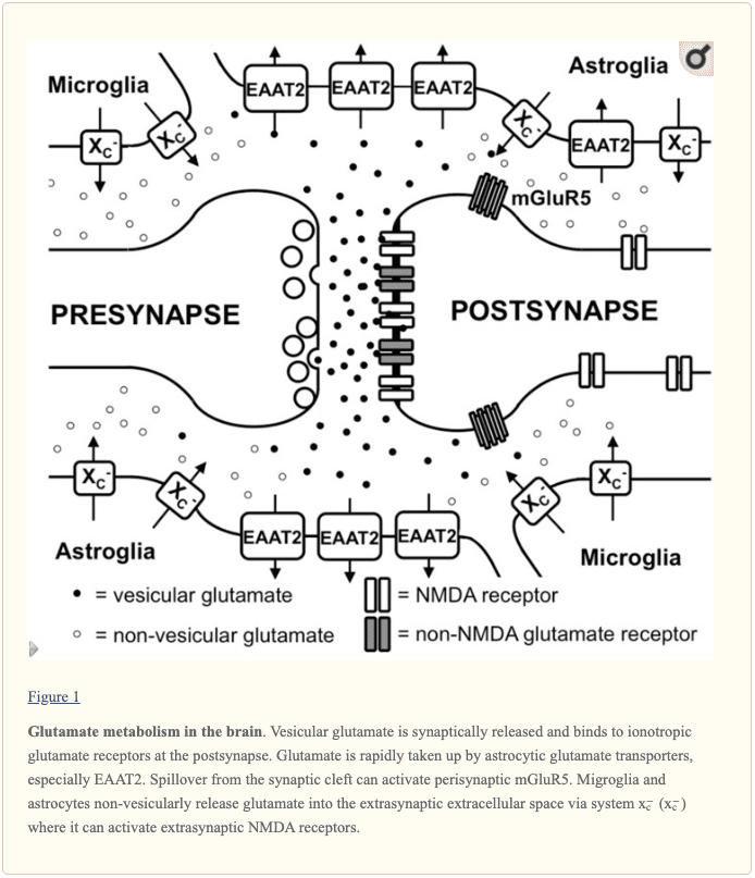
Additionally to iGluRs, there are also eight isoforms of metabotropic glutamate receptors (mGluRs) which belong to the family of G-protein-coupled receptors, where they don’t develop ion channels but instead signal through a variety of second messenger systems. L-glutamate-associated depolarization causes a postsynaptic excitatory potential which eases the development of an action potential at the axon hillock. The glutamatergic synapse is activated by astrocytic processes that demonstrate high levels of excitatory amino acid transporters (EAATs). There are five different EAATs, EAAT1 to 5, of which EAAT1 and 2 are the primary astrocytic EAATs, whereas EAAT3 shows a predominantly neuronal expression. Approximately 90 percent of the L-glutamate transport is regulated and maintained by EAAT2 such as GLT-1 in rodent models. These transporters then co-transport 2 or 3 molecules of Na+ and a proton with each molecule of L-glutamate or L-aspartate together with the counter-transport of a K+ ion. Therefore, by utilizing the electrochemical gradient of these ions throughout the plasma membrane as an energy source, the transporters are able to safely and effectively accumulate L-glutamate and L-aspartate in cells against their sudden intra- to extracellular concentration gradients. This allows the brain to control a very low extracellular L-glutamate concentration in the low micromolar range. It is generally believed that L-glutamate taken up by astrocytes is turned to glutamine by the enzyme glutamine synthetase, the glutamine is then released, taken up by neurons and turned to L-glutamate, where it is ultimately utilized once again for neurotransmission. �
Extrasynaptic Glutamate in the Brain
Aside from the essential role of L-glutamate as the primary excitatory neurotransmitter released from glutamatergic presynapses, as previously mentioned above, it has become evident that L-glutamate receptors outside the synaptic cleft also play an essential role in brain physiology. In the cerebellum, it was demonstrated by evaluating AMPA receptor-mediated currents in Bergmann glia that synaptically released L-glutamate concentrations can reach extrasynaptic concentrations of up to 190 ?M while concentrations in the synaptic cleft can exceed 1 mM. Moreover, several mGluRs have been shown to demonstrate a different localization in proximity to the postsynaptic density which would allow them to immediately recognize L-glutamate escaping from the synaptic cleft, as shown in Figure 1. However, current research studies have demonstrated that iGluRs, especially of the NMDA type, are also found at extrasynaptic regions in the neuronal cell membrane. Utilizing light and electron microscopy, other research studies also demonstrated that extrasynaptic NMDA receptors gather at different regions of close contact in the dendritic shaft with axons, axon terminals, or astrocytic processes. The proportion of extrasynaptic NMDA receptors was estimated to be as high as 36 percent of the dendritic NMDA receptor pool in rat hippocampal slices. Although extrasynaptic NMDA receptors were associated with similar scaffolding proteins as synaptic NMDA receptors, an in vitro research study suggested that extrasynaptic and synaptic NMDA receptors may ultimately activate different downstream signaling pathways with a variety of results, including the suppression of CREB activity by extrasynaptic NMDA receptor activation as well as activation by synaptic NMDA receptors. Furthermore, NMDA receptors localized extrasynaptically on dendritic shafts connect extrasynaptic L-glutamate as well as regulate and maintain Ca2+ influx during the elimination of the Mg+2 block by dendrite depolarization throughout the backfiring of action potentials. Research studies demonstrated that L-glutamate release from astrocytes can activate slow inward currents through extrasynaptic NMDAR receptors in CA1 neurons which can also be ultimately synchronized. The mechanisms through which glial cells release L-glutamate as well as how the extrasynaptic L-glutamate concentrations are controlled are vital towards understanding how the activity of extrasynaptic NMDA receptors is controlled. �
Different mechanisms through which astrocytes can release L-glutamate have been suggested, including vesicular L-glutamate release and non-vesicular release through anion channels as well as connexin hemichannels and release through the cystine/glutamate antiporter system x?c. Several research studies strongly suggest that vesicular release from astrocytes plays a minor role because the Ca+2-associated release of L-glutamate was still present in astrocytes created from dominant-negative SNARE mice where vesicular release can be blocked by doxycycline withdrawal. System x?c is a cystine/glutamate antiporter which is characterized as heterodimeric amino acid transporters, made up of xCT as the specific subunit and 4F2hc as the promiscuous heavy chain. This transporter is demonstrated in the brain, especially in astroglial and microglial cells, as shown in Figure 1. The fact that extrasynaptic L-glutamate levels in different regions of the human brain are downregulated by approximately 60 percent to 70 percent in xCT knock out mice, research studies demonstrated that system x?c releases L-glutamate into the extrasynaptic space and suggests that this transporter is essential in the regulation of extrasynaptic L-glutamate levels. This is further supported by the observation that when measured by in vivo microdialysis, the increase in extrasynaptic L-glutamate developed by EAAT inhibitors is neutralized by blocking system x?c while blocking neuronal vesicular L-glutamate release is ineffective. Further research studies are still required. �
Taken together, glutamatergic neurotransmissions don’t simply happen through classical excitatory synapses but also through extrasynaptic L-glutamate receptors, as shown in Figure 1. Finally, the levels of extrasynaptic L-glutamate are determined, at least partially, by glial non-vesicular L-glutamate release, as also shown in Figure 1. However, the regulation of extrasynaptic L-glutamate levels, as well as its temporal-spatial dynamics and its effect on neuronal function, neurodegeneration, and behavior, are far from being fully understood by researchers, healthcare professionals, and patients. �

Glutamate, together with aspartate, is one of the main excitatory neurotransmitters in the human brain. Although it plays a fundamental role in the overall structure and function of the nervous system, excessive amounts of glutamate can ultimately cause excitotoxicity which may lead to a variety of health issues, such as Alzheimer’s disease and other types of neurological diseases. The following article describes the role of glutamate in the human brain. – Dr. Alex Jimenez D.C., C.C.S.T. Insight
L-glutamate is one of the main excitatory neurotransmitters in the human brain and it plays an essential role in practically all activities of the nervous system. In the article above, we discussed the general principles of L-glutamate signaling in the brain. Then, we demonstrated this scheme by describing the different pools of extracellular glutamate, including the synaptic, the perisynaptic, and the extrasynaptic, resulting from vesicular and non-vesicular sources or abnormally located glutamate receptors outside of synapses as well as discussed their possible physiological functions in the human brain. The scope of our information is limited to chiropractic, musculoskeletal and nervous health issues as well as functional medicine articles, topics, and discussions. We use functional health protocols to treat injuries or chronic disorders of the musculoskeletal system. To further discuss the subject matter above, please feel free to ask Dr. Alex Jimenez or contact us at 915-850-0900 . �
Curated by Dr. Alex Jimenez �
References �
- Lewerenz, Jan, and Pamela Maher. �Chronic Glutamate Toxicity in Neurodegenerative Diseases-What Is the Evidence?� Frontiers in Neuroscience, Frontiers Media S.A., 16 Dec. 2015, www.ncbi.nlm.nih.gov/pmc/articles/PMC4679930/.
Additional Topic Discussion: Chronic Pain
Sudden pain is a natural response of the nervous system which helps to demonstrate possible injury. By way of instance, pain signals travel from an injured region through the nerves and spinal cord to the brain. Pain is generally less severe as the injury heals, however, chronic pain is different than the average type of pain. With chronic pain, the human body will continue sending pain signals to the brain, regardless if the injury has healed. Chronic pain can last for several weeks to even several years. Chronic pain can tremendously affect a patient’s mobility and it can reduce flexibility, strength, and endurance.
Neural Zoomer Plus for Neurological Disease
 �
�
Dr. Alex Jimenez utilizes a series of tests to help evaluate neurological diseases. The Neural ZoomerTM Plus is an array of neurological autoantibodies which offers specific antibody-to-antigen recognition. The Vibrant Neural ZoomerTM Plus is designed to assess an individual�s reactivity to 48 neurological antigens with connections to a variety of neurologically related diseases. The Vibrant Neural ZoomerTM Plus aims to reduce neurological conditions by empowering patients and physicians with a vital resource for early risk detection and an enhanced focus on personalized primary prevention. �
Formulas for Methylation Support

XYMOGEN�s Exclusive Professional Formulas are available through select licensed health care professionals. The internet sale and discounting of XYMOGEN formulas are strictly prohibited.
Proudly,�Dr. Alexander Jimenez makes XYMOGEN formulas available only to patients under our care.
Please call our office in order for us to assign a doctor consultation for immediate access.
If you are a patient of Injury Medical & Chiropractic�Clinic, you may inquire about XYMOGEN by calling 915-850-0900.
![]()
For your convenience and review of the XYMOGEN products please review the following link.*XYMOGEN-Catalog-Download �
* All of the above XYMOGEN policies remain strictly in force.
�
General Disclaimer, Licenses and Board Certifications *
Professional Scope of Practice *
The information herein on "Functional Neurology: The Role of Glutamate in the Brain" is not intended to replace a one-on-one relationship with a qualified health care professional or licensed physician and is not medical advice. We encourage you to make healthcare decisions based on your research and partnership with a qualified healthcare professional.
Blog Information & Scope Discussions
Welcome to El Paso's Premier Wellness and Injury Care Clinic & Wellness Blog, where Dr. Alex Jimenez, DC, FNP-C, a Multi-State board-certified Family Practice Nurse Practitioner (FNP-BC) and Chiropractor (DC), presents insights on how our multidisciplinary team is dedicated to holistic healing and personalized care. Our practice aligns with evidence-based treatment protocols inspired by integrative medicine principles, similar to those on this site and on our family practice-based chiromed.com site, focusing on naturally restoring health for patients of all ages.
Our areas of multidisciplinary practice include Wellness & Nutrition, Chronic Pain, Personal Injury, Auto Accident Care, Work Injuries, Back Injury, Low Back Pain, Neck Pain, Migraine Headaches, Sports Injuries, Severe Sciatica, Scoliosis, Complex Herniated Discs, Fibromyalgia, Chronic Pain, Complex Injuries, Stress Management, Functional Medicine Treatments, and in-scope care protocols.
Our information scope is multidisciplinary, focusing on musculoskeletal and physical medicine; wellness; contributing etiological viscerosomatic disturbances within clinical presentations; associated somato-visceral reflex clinical dynamics; subluxation complexes; sensitive health issues; and functional medicine articles, topics, and discussions.
We provide and present clinical collaboration with specialists from various disciplines. Each specialist is governed by their professional scope of practice and licensure jurisdiction. We use functional health & wellness protocols to treat and support care for musculoskeletal injuries or disorders.
Our videos, posts, topics, and insights address clinical matters and issues that directly or indirectly relate to our clinical scope of practice.
Our office has made a reasonable effort to provide supportive citations and has identified relevant research studies that support our posts. We provide copies of supporting research studies upon request to regulatory boards and the public.
We understand that we cover matters that require an additional explanation of how they may assist in a particular care plan or treatment protocol; therefore, to discuss the subject matter above further, please feel free to ask Dr. Alex Jimenez, DC, APRN, FNP-BC, or contact us at 915-850-0900.
We are here to help you and your family.
Blessings
Dr. Alex Jimenez, DC, MSACP, APRN, FNP-BC*, CCST, IFMCP, CFMP, ATN
email: [email protected]
Multidisciplinary Licensing & Board Certifications:
Licensed as a Doctor of Chiropractic (DC) in Texas & New Mexico*
Texas DC License #: TX5807, Verified: TX5807
New Mexico DC License #: NM-DC2182, Verified: NM-DC2182
Multi-State Advanced Practice Registered Nurse (APRN*) in Texas & Multi-States
Multi-state Compact APRN License by Endorsement (42 States)
Texas APRN License #: 1191402, Verified: 1191402 *
Florida APRN License #: 11043890, Verified: APRN11043890 *
Colorado License #: C-APN.0105610-C-NP, Verified: C-APN.0105610-C-NP
New York License #: N25929, Verified N25929
License Verification Link: Nursys License Verifier
* Prescriptive Authority Authorized
ANCC FNP-BC: Board Certified Nurse Practitioner*
Compact Status: Multi-State License: Authorized to Practice in 40 States*
Graduate with Honors: ICHS: MSN-FNP (Family Nurse Practitioner Program)
Degree Granted. Master's in Family Practice MSN Diploma (Cum Laude)
Dr. Alex Jimenez, DC, APRN, FNP-BC*, CFMP, IFMCP, ATN, CCST
(Board Certified: Family Practice Nurse Practitioner—Multistate)*
(Licensed Nurse Practitioner & Chiropractor - Multistate)*
Clinical Director
Digital Business Card
Dr. Maria Cardenas, MD
(Board Certified: Internal Medicine)
(Licensed Medical Doctor)
Medical Director, Clinical Director & Collaborative Physician
NPI # 1164426749
MD License #: J2933
Licenses and Board Certifications:
MD: Medical Doctor
DC: Doctor of Chiropractic
APRNP: Advanced Practice Registered Nurse
FNP-BC: Family Practice Specialization (Multi-State Board Certified)
RN: Registered Nurse (Multi-State Compact License)
CFMP: Certified Functional Medicine Provider
MSN-FNP: Master of Science in Family Practice Medicine
MSACP: Master of Science in Advanced Clinical Practice
IFMCP: Institute of Functional Medicine
CCST: Certified Chiropractic Spinal Trauma
ATN: Advanced Translational Neutrogenomics
Memberships & Associations:
TCA: Texas Chiropractic Association: Member ID: 104311
AANP: American Association of Nurse Practitioners: Member ID: 2198960
ANA: American Nurse Association: Member ID: 06458222 (District TX01)
TNA: Texas Nurse Association: Member ID: 06458222
NPI: 1205907805
| Primary Taxonomy | Selected Taxonomy | State | License Number |
|---|---|---|---|
| No | 111N00000X - Chiropractor | NM | DC2182 |
| Yes | 111N00000X - Chiropractor | TX | DC5807 |
| Yes | 363LF0000X - Nurse Practitioner - Family | TX | 1191402 |
| Yes | 363LF0000X - Nurse Practitioner - Family | FL | 11043890 |
| Yes | 363LF0000X - Nurse Practitioner - Family | CO | C-APN.0105610-C-NP |
| Yes | 363LF0000X - Nurse Practitioner - Family | NY | N25929 |
Dr. Alex Jimenez, DC, APRN, FNP-BC*, CFMP, IFMCP, ATN, CCST
(Board Certified: Family Practice Nurse Practitioner—Multistate)*
(Licensed Nurse Practitioner & Chiropractor - Multistate)*
Clinical Director
Digital Business Card
Dr. Maria Cardenas, MD
(Board Certified: Internal Medicine)*
(Licensed Medical Doctor)*
Medical Director, Clinical Director & Collaborative Physician
NPI # 1164426749
MD License #: J2933


